 |
| |
||
| |
|||||||||||||||||
|
|||||||||||||||||
|
|||||||||||||||||
| EID
Home | Ahead of Print | Past
Issues | EID Search | Contact
Us | Announcements | Suggested
Citation | Submit Manuscript
Volume 10, Number 11, November 2004 Topographic Changes in SARS Coronavirus–infected Cells at Late Stages of InfectionM.L. Ng,* J.W.M. Lee,* M.L.N. Leong,* A.-E. Ling,† H.-C. Tan‡ and
E.E. Ooi‡ |
||
|
|
  |
|
| Back to article | |
|
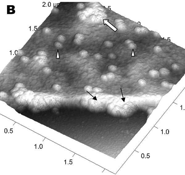
Figure 6. Atomic force microscopy of Vero cells infected with severe acute respiratory syndrome–associated coronavirus. A) High activity of virus extrusion at the thickened edge of the infected cells (arrow). Arrowheads indicate virus particles. B) A three-dimensional reconstruction of the image in panel A shows the puffy edge of infected cells. Many intracellular viruses are visible just under the plasma membrane (arrows). Extruded virus particles are present on other areas of the cell surface (arrowheads). Thick white arrow shows a large clump of virus particles just beneath the plasma membrane. |
|
|
|
|
|
EID Home | Top of Page | Ahead-of-Print | Past Issues | Suggested Citation | EID Search | Contact Us | Accessibility | Privacy Policy Notice | CDC Home | CDC Search | Health Topics A-Z |
|
|
This page last reviewed October 12, 2004 |
|
|
Emerging
Infectious Diseases Journal |
|