Trichomonas vaginalis Parasite
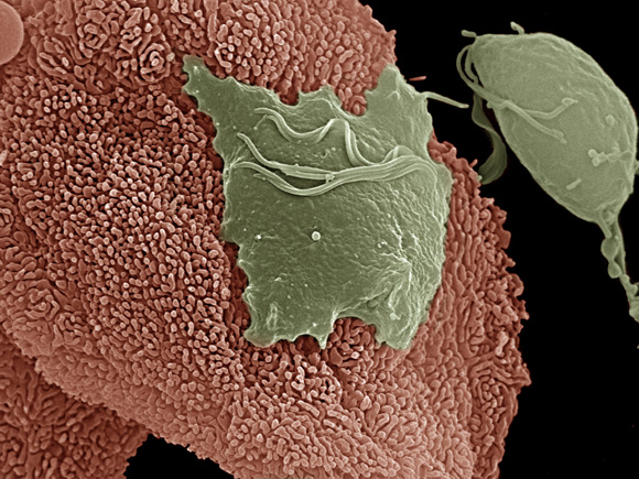
An electron micrograph depicts the Trichomonas vaginalis parasite adhering to vaginal epithelial cells collected from vaginal swabs. A non-adhered parasite (right) is pear-shaped, whereas the attached parasite is flat and amoeboid.
Image courtesy of: Antonio Pereira-Neves and Marlene Benchimol, Santa Ursula University, Rio de Janeiro, Brazil
Return to News Release
back to top