Laboratory Personnel, Slide 3
Comparison of Hemolytic Activity for Group A, B, G, F Strep
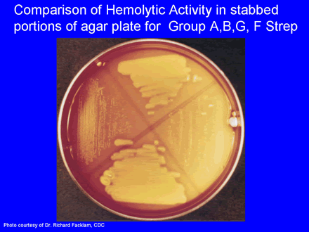
This is a picture of another blood agar plate. In this picture, the group A Streptococcus is in the quadrant on the top portion of the agar plate; the group B Streptococcus is shown on the right quadrant; a group G Streptococcus is shown on the bottom quadrant; and a group F Streptococcus is shown on the left quadrant. Although the differences may not be apparent to the untrained eye, there are small differences in the size of the hemolytic activity of each of these different streptococci, especially in the stabbed portion of the agar plate as depicted by the arrows. The GBS is the bacteria that causes the least amount of hemolytic activity. And the group F Streptococcus has the smallest colony size.
Page Last Modified: April 20, 2008
Content Last Reviewed: April 20, 2008
Content Source: National Center for Immunization and Respiratory Diseases