|
|
Diagnosis
Asbestos-related conditions can be difficult to identify. Healthcare providers usually identify the possibility of asbestos exposure and related health conditions, like lung and pleural disease, by taking a thorough medical history. This includes looking at the person's medical, work, cultural, and environmental history. After a physician suspects an asbestos-related health condition, he or she can use a number of tools to help make the actual diagnosis.
An accurate medical history is essential for an accurate diagnosis.
Ways to Detect Lung and Pleural Disease include
- Physical Examination
- Chest X-ray
- Pulmonary function tests
- Biopsy/Bronchoscopy
- Computed Tomography Scans
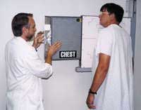
1. Physical Examination
2. Chest X-ray - The chest X-ray is the most common tool used to detect lung and pleural disease caused by chronic exposure to asbestos. X-rays often show pleural changes in people who have been exposed to asbestos. Although X-rays cannot specifically determine if a condition is asbestos-related, lung abnormalities can be noted. These changes include pleural plaques (the most frequent lesions): discrete, elevated, opaque, shiny fibrosis lesions that are currently more common than asbestosis in exposed persons; diffuse pleural thickening and pleural effusions, are early manifestations of inhalation exposure to high concentrations of asbestos. Pleural effusions can be an early indication of mesothelioma and warrant further evaluation. Early identification of mesothelioma and intervention can increase chances of survival
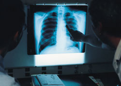
Chest X-Rays are the most common method of detecting asbestos-related disorders.
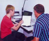
3. Pulmonary function tests
A patient performs a pulmonary function test.
4. Biopsy/Bronchoscopy - A lung biopsy to detect asbestos fibers in pieces of lung tissue, while not needed to make a clinical diagnosis, is the most reliable test to determine the presence of asbestos-related effects. Less invasive tests (e.g. bronchoscopy) can detect asbestos fibers or asbestos bodies in bronchoalveolar lavage fluid or in sputum. These tests, however, do not reliably indicate how much asbestos a person may have been exposed to or predict whether disease will develop.
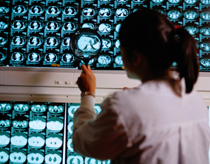
5. Computed Tomography Scans (CT Scans) - Over the past 20 years, several investigators have evaluated the potential of conventional scans for detecting abnormalities in asbestos-exposed workers. During 2001, ATSDR completed a study examining the effectiveness of CT-scans in detecting asbestos-related lung abnormalities as a part of a medical testing program conducted in Libby, Montana, where asbestos exposure occurred. Click here to read the protocol for the ATSDR study. A CT scan is an image produced by an X-ray source that rotates around the patient. The beams pass through the patient and are detected on the other side. This information is analyzed by computer to generate a cross-sectional view of the patient at that level. Studies have shown that CT offers several advantages over routine chest radiography (X-ray). First, the use of cross-sectional images often makes it possible to distinguish between densities that would be superimposed on plain radiographs. Second, CT is far better than routine radiographic studies at characterizing tissue density, distinguishing subtle differences in density between adjacent structures, and providing accurate size assessment of lesions.
Treatment for Asbestos-Related Diseases
Treatment for Nonmalignant-Illnesses
Unfortunately, no cure exists for asbestosis or other lung or pleural diseases caused by chronic exposure to asbestos. Treatment involves preventing further complications and treating symptoms.
Preventing further exposure and ceasing any tobacco smoking activities are the most important steps individuals can take to minimize development of health problems. Once established, these diseases can remain stable or progress in severity in the absence of further exposure. The diseases rarely regress.
Treatment for Malignant Illnesses
Treatment options for patients diagnosed with asbestos-related cancer of the lung or pleura are limited to resection (surgical removal of a part), chemotherapy, or both.
Future research could help to develop therapeutic methods to interfere with the development of asbestos-induced lung and pleural disorders to cause the disorders to regress once they are established.
The National Jewish Medical and Research Center![]() partnered with ATSDR in its asbestos investigations in Libby, Montana. The center treats asbestos-related illnesses. To learn more about the center and how to access its services, visit their Web site
partnered with ATSDR in its asbestos investigations in Libby, Montana. The center treats asbestos-related illnesses. To learn more about the center and how to access its services, visit their Web site![]() .
.
The National Cancer Institute![]() web site also offers more information on treatment.
web site also offers more information on treatment.
Recommendations
The following are important steps for minimizing risks of asbestos-related disorders:
- Minimize or avoid further exposure to any form of asbestos;
- Stop smoking and avoid tobacco smoke (second hand) and other pulmonary sensitizers and irritants;
- Get regular medical care;
- Consider appropriate vaccines (such as influenza and pneumonia) based on physician recommendations to prevent other pulmonary infections.
ATSDR expects people who were exposed to asbestos and who smoke to be unusually susceptible to asbestos-related lung cancer and asbestosis. Studies of asbestos workers indicate that asbestos-exposed smokers have greater than additive risks for lung cancer and asbestosis than asbestos-exposed nonsmokers. These people are strongly encouraged to quit smoking. See the patient handout Cigarette Smoking, Asbestos Exposure, and your Health.
Individuals living or working in buildings with vermiculite insulation or other building materials that could contain asbestos are encouraged to ensure that the insulation material is in good repair and not disturbed in order to prevent the release of asbestos fibers. If the material is to be removed or disturbed for any reason, special procedures to minimize the generation of dust and specify appropriate locations for disposal must be followed. Individuals can obtain information about asbestos removal and disposal procedures from the 10 regional offices of the EPA![]() .
.
Medical Testing Information
ATSDR Medical Testing In Libby, Montana
ATSDR conducted face-to-face interviews with individuals to help determine the extent of asbestos-related illness in Libby, Montana.
This medical testing program was designed to
- Identify asbestos-related health effects among people exposed to asbestos from the Libby vermiculite mine and refer them for additional evaluation and treatment
- Provide the EPA with information needed to identify and eliminate any significant exposures to asbestos in the community
- Identify the types of illnesses experienced by these exposed people in order to better educate local physicians
- Provide the local medical community with an estimate of the additional medical care the community will need over the next 10-20 years. This plan was developed by ATSDR in close collaboration with other federal, state, and local agencies.
Participants were encouraged to consult their individual health care providers. ATSDR does not provide and is not authorized by Congress to provide medical diagnosis or treatment.
The medical testing consisted of (1) a face-to-face questionnaire designed to obtain exposure information, including detailed information about potential pathways of exposure and to collect information about respiratory symptoms, demographics, and smoking; (2) spirometry to provide an objective measurement of lung function and (3) chest radiograph (X-ray) to identify asbestos-related changes in the parenchyma and pleura of the lungs. Chest X-rays were evaluated and were interpreted by onsite clinical radiologists as well as a panel of three national experts in asbestos-related conditions.
Right: A chest X-ray is the most common tool used to detect asbestos-related illnesses.
Results of Medical Testing
The results of the asbestos medical testing conducted the summer of 2000 are available (Read the Year 2000 Medical Testing for more details), and the results from the 2001 testing will be released in 2002. Major findings of the medical testing completed in 2000 include:
X-ray Findings
Experts found that 994 of the 5,590 participants - or 18% of those X-rayed - had abnormalities in the lining of their lungs (pleural abnormalities). The risk of pleural abnormality increased with increasing age and increasing length of residence in the Libby area. In comparison, the rate of pleural abnormalities found in groups within the U.S. who have no known asbestos exposures ranges from 0.2% to 2.3%.
- Forty-eight percent (159 of 328) of former W.R. Grace employees had pleural abnormalities.
- Most participants reported multiple routes of exposure (household contact, occupational, recreational, and other). For example, 24% of participants who reported six or more reported routes of exposure had pleural abnormalities.
- Five percent (6 of 122) of those participants who reported no apparent exposure had pleural abnormalities.
- Factors most strongly related to having pleural abnormalities were 1) having been a W.R. Grace/Zonolite worker, 2) having household contact with a W.R. Grace/Zonolite worker, and 3) being a male.
- The risk of finding a pleural abnormality is almost 8 times greater for former W.R. Grace workers when compared to non-W.R. Grace workers of the same age.
- The risk of finding a pleural abnormality is 3.3 times greater for females who have household contact with W.R. Grace workers when compared to women who have no household contacts with these workers.
- The risk of finding a pleural abnormality is 5 times greater for men than for women.
Lung Function Test Findings
Lung function tests (spirometry) were offered to all study participants. An on-site pulmonologist interpreted all tests. Some of the key findings are as follows:
- Being a current smoker was the strongest risk factor for restrictive changes.
- Moderate to severe restriction in breathing capacity was found in 5.7% of former W.R. Grace workers.
- Moderate to severe restriction in breathing capacity was found in 2.2% of men and 1.6% of women (men and women defined as 18 years of age and older).
- In those under 18 years of age who were tested, no one had moderate to severe restricted lung function.
- Other factors affecting restricted breathing included (a) being a non-W.R. Grace/Zonolite worker exposed to vermiculite, (b) having had chest surgery, and (c) being overweight.
Health Care Professionals Materials
Forms
- Taking an Exposure History
Print Version (0.14 MB)
This form helps health care professionals capture and evaluate the exposure history of their patients.
Quick Reference Guides
- Asbestos Disease: An Overview for Clinicians
Preview Version | Print Version (0.25 MB)
This brief overview describes the primary adverse effects of asbestos exposure, guidelines for assessing patient risk for asbestos-related disease, clinical evaluation techniques, and strategies for advising patients. - Clinical Screening Guidelines for Asbestos-Related Lung Disease
Preview Version | Print Version (0.19 MB)
The reference lists the steps to take in screening for asbestos-related lung disease: a medical history interview, performing a physical exam, conducting a PA chest radiograph, and performing a simple pulmonary function test. - ToxFAQs for Asbestos
HTML Version | Print Version (2.99 MB)
This is an abbreviated version of Toxicological Profile for Asbestos (see under Reference Guides) that gives an overview on the toxicological characteristics and health effects of asbestos. - Environmental Exposure History - "I Prepare" - Pocket Guide
Print Version (0.048 MB)
This is a quick reference card for conducting an exposure history.
Reference Guides
- Case Studies in Environmental Medicine: Taking an Exposure History
HTML Version
The goal of this 61-page case study is to increase the health care professional's knowledge of hazardous substances in the environment and to aid in the evaluation of potentially exposed patients. - Toxicological Profile for Asbestos
HTML Version | Print Version (2.99 MB)
This 441-page document gives information on the toxicological characteristics and health effects of asbestos.
Patient Materials
- Living with Asbestos-Related Illness [HTML] ****
Topics covered in the guide include characteristics of asbestos, asbestos-related illness, the respiratory system, treatment methods, preventive care, traveling tips, pulmonary rehabilitation, and relaxation and breathing techniques.
This page last updated on December 04, 2007
Agency for Toxic Substances and Disease Registry, 1825 Century Blvd, Atlanta, GA 30345
Contact CDC: 800-232-4636 / TTY: 888-232-6348