Skip Standard Navigation Links
Centers for Disease Control and Prevention
 CDC Home Search Health Topics A-Z
peer-reviewed.gif (582 bytes)
eid_header.gif (2942 bytes)
Past Issue

Vol. 12, No. 6
June 2006

Adobe Acrobat logo

EID Home | Ahead of Print | Past Issues | EID Search | Contact Us | Announcements | Suggested Citation | Submit Manuscript

PDF Version PDF Version | Comments Comments | Email this article Email this article


Materials and Methods
Results
Discussion
Acknowledgments
References
Figure 1
Figure 2
Figure 3
Table 1
Table 2

Research

Human Rotavirus Serotype G9, São Paulo, Brazil, 1996–2003

Rita Cássia Compagnoli Carmona,*Comments Maria do Carmo Sampaio Tavares Timenetsky,* Simone Guadagnucci Morillo,* and Leonardo José Richtzenhain†
*Adolfo Lutz Institute, São Paulo, Brazil; and †University of São Paulo, São Paulo, Brazil

Suggested citation for this article


A total of 3,101 fecal specimens were collected during an 8-year survey for rotavirus infection in São Paulo, Brazil. Group A rotavirus was detected in 774 (25.0%) specimens. Of these, 431 strains (55.7%) were analyzed for G and P types by reverse transcription–polymerase chain reaction; G1 was the predominant serotype (68.2%), followed by G9 (17.2%), G4 (6.3%), G2 (1.2%), G3 (0.7%), mixed infection (1.8%), and untypeable (4.6%). Both rotavirus G and P types could be established in 332 strains (77.0%). We identified the 4 most common strains worldwide: P[8]G1 (66.6%), P[4]G2 (1.0%), P[8]G3 (0.6%), and P[8]G4 (7.2%). Among the single G9 strains detected, VP4 genotyping showed that P[8]G9 was the most prevalent, followed by P[4]G9 and P[6]G9. The emergence and high frequency of rotavirus G9 in São Paulo, Brazil, and other parts of the world will affect the development and evaluation of future vaccines.

Group A rotavirus is the most common cause of acute gastroenteritis in infants and young children worldwide (1). More than 130 million cases of diarrhea each year are attributed to rotavirus. It is estimated to cause >400,000 deaths annually in children <5 years of age and is responsible for 2 million hospital admissions due to acute diarrhea worldwide. In developing countries, an estimated 1,205 children die from rotavirus disease each day, and 82% of these deaths occur in children in the poorest countries (2)

Rotavirus serotypes are determined by neutralizing antibody responses to each of the 2 outer capsids proteins, VP7 (G serotype) and VP4 (P serotype) (1). To date, 11 VP7 G serotypes and 13 P serotypes have been identified in humans. Serotypes G1, G2, G3, and G4 are frequently associated with diarrhea in humans and have become prime targets for vaccine development (3,4). The recent emergence and wide distribution of rotavirus G9 indicate that this serotype may become the fifth relevant strain (5,6). Unusual types of rotavirus have been described in certain settings. The G5 type has been reported in Brazil, Argentina, Paraguay, Cameroon, and the United Kingdom (6,7); G6 has been detected in Italy, Australia, India, the United States, Belgium, and Hungary; G8 has been frequently isolated in Africa and sporadically in other countries; rotavirus G10 specificity has been reported in the United Kingdom, India, Thailand, Paraguay, and Brazil (6); G11 type was recently detected in Dhaka, Bangladesh (4); and the G12 type has been detected in the Philippines (8), Thailand (9), the United States (10), India (11), Japan (12), Korea (13), Argentina (14), and Brazil (15).

The genotypes VP4 P[8] and P[4] are the most common P types that infect humans. The P[8] type is generally associated with VP7 types G1, G3, and G4, and the P[4] type is associated with G2 (16). Combined G and P genotyping may have advantages in identifying reassortants as unusual or new virus strains (17). Continued surveillance of the diverse rotavirus strains circulating in a community is crucial before developing a vaccine and during and after implementing an immunization program. Therefore, we describe the results of an 8-year surveillance study of G- and P-type rotavirus strains from persons with acute diarrhea in the state of São Paulo, Brazil.

Materials and Methods

Figure 1
Figure 1.

Click to view enlarged image

Figure 1. Map of São Paulo State, Brazil, indicating where fecal specimens were collected during the 8-year survey period.


Figure 2

Figure 2.
Click to view enlarged image

Figure 2. Temporal distribution of rotavirus strains from children, adults, and elderly patients with acute diarrhea, São Paulo, Brazil, 1996–2003.
  

Figure 3

Figure 3.
Click to view enlarged image

Figure 3. Rotavirus infection among children (<5 years of age), school-age children (5–17 years), adults (18–59 years)...

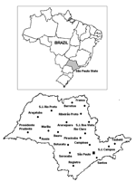
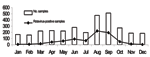
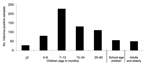
From 1996 to 2003, a total of 3,101 fecal specimens were collected from children <5 years of age, school-age children (5–17 years), adults (18–59 years), and elderly patients (>60 years) with acute gastroenteritis. These patients received treatment for diarrhea at the departments of public health or were admitted to hospitals in several cities in São Paulo State, in southeast Brazil. São Paulo State has an area of ≈248,800 km2 and a population of 40 million (21.5% of the population of Brazil). Figure 1 shows main cities in São Paulo where samples were collected. Epidemiologic data (age, date of diarrhea onset, date of sample collection) were available from some patients. Specimens were stored at –20°C until tested for rotavirus and characterized. Study methods were approved by the ethical committee of Adolfo Lutz Institute.

All specimens were screened for rotavirus by using a commercial enzyme-linked immunosorbent assay (ELISA) (Premier Rotaclone, Meridian Diagnostics, Cincinnati, OH, USA) with monoclonal antibodies specific for group A human rotavirus, according to the manufacturer's protocol. Rotavirus double-stranded RNA (dsRNA) was extracted directly from stool by the TRIzol method (Invitrogen, Carlsbad, CA, USA) and precipitated with isopropanol. The extracted dsRNA was subjected to G and P typing by multiplex reverse transcription–polymerase chain reaction (RT-PCR) with type-specific primers. Consensus primers Beg9 and End9 were used in a first-round PCR (30 cycles) to amplify the full-length VP7 gene (1,062 bp); cDNA was used in a second-round PCR for G typing (25 cycles) with primer set aBT1 (G1), aCT2 (G2), aET3 (G3), aDT4 (G4), aFT9 (G9) and primer set FT5 (G5), DT6 (G6), HT8 (G8), ET10 (G10), BT11 (G11) (18,19). For P typing, consensus primers Con2 and Con3 were used in a first-round RT-PCR (30 cycles) to amplify the 876 bp of the VP8* region of the VP4 gene, and the second-round PCR (20 cycles) used primer set 1T-1 (P[8]), 2T-1 (P[4]), 3T-1 (P[6]), 4T-1 (P[9]), 5T-1 (P[10]) (20). All PCR products were analyzed by electrophoresis in 1.2% agarose gels, containing 0.5 µg ethidium bromide per milliliter and visualized under UV illumination.

Results

Rotavirus was detected in 774 (25.0%) of 3,101 specimens collected from children, adults, and elderly patients in São Paulo during an 8-year period. Rotavirus infection was found predominantly in the winter and in drier months. The incidence peaked in August (Figure 2). The age or date of birth was provided for 677 (87.5%) of 774 patients who tested positive for rotavirus. Rotavirus disease was detected mainly in children <2 years of age (463 [59.8%] of 774), and peaks of incidence occurred from 7 to 12 months (Figure 3). However, rotavirus infection also was detected in adults and elderly patients (55 [7.1%] of 774).

We randomly selected 431 rotavirus-positive samples (55.7%) for determination of G and P genotypes by an RT-PCR assay. G1 was the predominant serotype in these samples (294, 68.2%), followed by G9 (74, 17.2%), G4 (27, 6.3%), G2 (5, 1.2%), G3 (3, 0.7%), mixed infection (8, 1.8%), and untypeable (20, 4.6%) (Table 1). The distribution of rotavirus types in São Paulo during this 8-year period shows that G1 was the most prevalent genotype in most years, but it was displaced by G9 in 2002. Incidence of G2 and G3 serotypes was low during the period of analysis. Frequency of G4 serotype differed during the surveillance period; it was not detected in 1996 and 2003. We found several mixed infections from 2000 to 2003 (G1+G4, G1+G9, G2+G3, and G4+G9).

Both rotavirus G and P types could be established in 332 (77.0%) strains, and 17 different P and G associations were detected (Table 2). Of these, we identified the 4 most globally common strains, P[8]G1, P[8]G4, P[4]G2, and P[8]G3, which represented 75.3% of all typed rotavirus strains. Uncommon strains were also detected, including P[8]G9, P[4]G9, P[6]G9, P[4]G1, P[6]G1, P[6]G2, and P[4]G4. And combination P-G mixed infections were as diverse as P[8]G1+G4, P[8]G1+G9, P[6]G1+G9, P[4]G2+G3, and P[8]G4+G9.

Discussion

We detected rotaviruses in the specimens of 25.0% of patients with acute diarrhea, which is comparable to the prevalence seen in other studies in Brazil (21). Among children <5 years of age, we detected rotavirus infection mainly in those <2 years of age (81.0%) (data not shown); in adults, rotavirus was detected less frequently (7.1%). The finding of a low percentage of rotavirus infection among adults is likely because the disease is generally perceived to be a childhood infection (22). Common epidemiologic settings for rotavirus infection among adults include endemic disease, epidemic outbreak, travel-related infection, and child-to-adult transmission (23).

In studies performed at various locations in Brazil with diverse climatic conditions, rotavirus disease appears to occur year-round (24). In São Paulo State, however, infection occurred mainly during cooler and drier seasons; similar observations have been made in other countries with temperate climates (16).

During the 8-year period studied, the G1 type was the most prevalent rotavirus strain. The second most prevalent was the G9 type, which accounted for 17.2% of disease, followed by G4, G2, and G3, which are common around the world. G1 was the most prevalent type in most years; however, it was displaced by G9 during the 2002 season, when G9 accounted for 46.9% of typed isolates. The G9 type has been reported to be a common cause of diarrhea and has become the fifth most common serotype, which suggests that it may be a substantial cause of diarrhea in humans (5,6). This type has been detected in Brazil since 1997 (25,26).

Surveillance on rotavirus types has been performed in São Paulo for >18 years, from 1986 to 2003 (27,28). The first G9 type was isolated in 2000 and has been fluctuating in frequency since its emergence (Table 1). The G5 type, normally associated with animal rotavirus (pigs and horses), has been frequently detected in persons in Brazil and it was considered an endemic virus (28,29). Nevertheless, in our survey in São Paulo, G5 rotavirus was not detected. Its incidence in Brazil has been decreasing over the last few years, and it may be disappearing (25,26); this type of rotavirus is likely to be a cyclic form. G9 strains have also been detected in animals (lambs and pigs [1]); detection of animal rotavirus provides evidence for natural human-animal genetic reassortment (30). Surveillance programs for animal rotavirus may aid in the development of next-generation vaccines (6).

Characterization of rotavirus VP4 types showed various strains. In this study, the 4 most globally common strains, P[8]G1, P[4]G2, P[8]G3, and P[8]G4, represented 75.3% of all typed viruses. The most prevalent association was P[8]G1, followed by P[8]G9. Worldwide, the 4 predominant rotavirus genotypes make up nearly 90% of all rotavirus infections (16). In Brazil, epidemiologic data on the prevalence of G and P types have been collected since the 1980s (27). This study showed great diversity of rotavirus strains in São Paulo. The uncommon genotypes P[4]G1, P[4]G4, P[4]+P[6]G1, and P[6]+P[8]G1 were also seen in some cases, similar to results from other countries (31,32). In our study, the uncommon genotypes P[6]G1, P[6]G2, and P[6]G9 were detected in children with acute diarrhea with an average age of 14 months (data not shown). Many studies have shown that this type is often detected in very young children with diarrhea, which suggests that P[6] strains may promote infection at an early age. Originally, P[6] in association with rotavirus types G1–G4 was detected in asymptomatic neonates (33), but recent studies have also shown that P[6]G9 circulates in hospitalized children without diarrhea (34). These strains are considered naturally attenuated and have been used to develop a vaccine candidate. Worldwide, rotavirus strains with the P[6] genotype have been seen in children with diarrhea (35–37). In Brazil, other studies isolated the P[6] type from children with acute diarrhea (26–28). In our survey, several mixed G and P types also appeared but in a low percentage (2.6%). The detection of unusual strains and mixed infections in this study suggests a previously unrecognized diversity among Brazilian rotavirus infections (28).

Among the single G9 strains detected in this survey, VP4 genotyping showed that P[8]G9 was the most prevalent (75.7%), followed by P[4]G9 (5.4%) and P[6]G9 (1.4%) (data not shown). This diversity among G9 types has also been detected in other studies (5,6,26). Combined data from P and G typing are relevant to identify new strains that might have resulted from reassortment of genes between diverse human-human and human-animal rotaviruses (38).

Recently, 2 live rotavirus oral vaccines have been licensed in some countries and made available on the market, including a monovalent vaccine derived from the most common human rotavirus strain, P[8]G1, and a pentavalent vaccine based on a bovine strain, WC3, that contains 5 human-bovine reassortant viruses (G1, G2, G3, G4, and P[8]). Both vaccines have shown efficacy against severe rotavirus disease (39,40). In August 2005, the live, attenuated P[8]G1 human rotavirus vaccine was licensed in Brazil, the first country to introduce this vaccine into the public health network.

Our data show that challenges exist for the design of rotavirus vaccine for the Brazilian population and underscore that virus strain surveillance should be ongoing. Surveillance programs can establish whether G9 rotavirus strains will continue to rise in prevalence or whether they will follow a cyclical pattern of emergence, as has been shown for G1–G4. The composition of future rotavirus vaccines is likely to be formulated according to the geographic setting and the distribution of G and P strains.

Acknowledgments

We thank Carla Uchida Tanuma, Daniela Bernardes Borges, Fernanda Ferreira da Silva, Karla Tatiana Rubini, Raquel de Souza Pereira, and Tomoko Sekiya for their technical assistance.

This work was supported by Adolfo Lutz Institute, São Paulo State Secretary of Health, São Paulo.

Dr Carmona is a research scientist in the Enteric Virus Laboratory, Adolfo Lutz Institute, São Paulo State Department of Health, São Paulo, Brazil. Her research interests are focused on the diagnosis of enteric virus diseases and the molecular characterization and epidemiology of rotavirus, norovirus, and enteroviruses.

References

  1. Kapikian AZ, Hoshino Y, Chanock RM. Rotaviruses. In: Knipe DM, Howley PM, Griffin DE, Martin MA, Lamb RA, Roizman B, et al., editors. Fields virology. Volume 2. 4th ed. Philadelphia: Lippincott-Raven; 2001. p. 1787–833.
  2. Parashar UD, Gibson CJ, Breese JS, Glass RI. Rotavirus and severe childhood diarrhea. Emerg Infect Dis. 2006;12:304–6
  3. Hoshino Y, Kapikian AZ. Rotavirus serotypes: classification and importance in epidemiology, immunity, and vaccine development. J Health Popul Nutr. 2000;18:5–14
  4. Rahman M, Matthijnssens J, Nahar S, Podder G, Sack DA, Azim T, et al. Characterization of a novel P[25], G11 human group a rotavirus. J Clin Microbiol. 2005;43:3208–12.
  5. Castello AA, Arvay ML, Glass RI, Gentsch J. Rotavirus strain surveillance in Latin America. A review of the last nine years. Pediatr Infect Dis J. 2004;23:S168–72.
  6. Santos N, Hoshino Y. Global distribution of rotavirus serotypes/genotypes and its implication for the development and implementation of an effective rotavirus vaccine. Rev Med Virol. 2005;15:29–56.
  7. Beards G, Graham C. Temporal distribution of rotavirus G-serotypes in the West Midlands region of the United Kingdom, 1983–1994. J Diarrhoeal Dis Res. 1995;13:235–7.
  8. Taniguchi K, Urasawa T, Kobayashi N, Gorziglia M, Urasawa S. Nucleotide sequence of VP4 and VP7 genes of human rotaviruses with subgroup I specificity and long RNA pattern: implication for new serotype specificity. J Virol. 1990;64:5640–4.
  9. Pongsuwanna Y, Guntapong R, Chiwakul M, Tacharoenmuang R, Onvimala N, Wakuda M, et al. Detection of a human rotavirus with G12 and P[9] specificity in Thailand. J Clin Microbiol. 2002;40:1390–4.
  10. Griffin DD, Nakagomi T, Hoshino Y, Nakagomi O, Kirkwood CD, Parashar UD, et al. Characterization of nontypeable rotavirus strains from the United Status: identification of a new rotavirus reassortant (P2A[6], G12) and rare P3[9] strains related to bovine rotavirus. Virology. 2002;294:256–69.
  11. Das S, Varghese V, Chaudhury S, Barman P, Mahapatra S, Kojima K, et al. Emergence of novel human group A rotavirus G12 strain in India. J Clin Microbiol. 2003;41:2760–2.
  12. Shinozaki K, Okada M, Nagashima S, Kaiho I, Taniguchi K. Characterization of human rotavirus strains with G12 and P[9] detected in Japan. J Med Virol. 2004;73:612–6.
  13. Cheon DS, Lee K, Kim W, Lee S, Choi W, Ahn J, et al. Genetic analysis of the VP7 gene of unusual genotypes of human group A rotavirus strains circulating in Korea [abstract P37-3]. In: Abstracts of the 23rd Annual Meeting of the American Society for Virology; Montreal, Canada; 2004 Jul 10–14. American Society for Virology; 2004.
  14. Castello AA, Jiang B, Glass RI, Glikmann G, Gentsch JR. Rotavirus G and P genotype prevalence in Argentina 1999–2003. Detection of P[9]G12 strains [abstract P37-4]. In: Abstracts of the 23rd Annual Meeting of the American Society for Virology; Montreal, Canada; 2004 Jul 10–14. American Society for Virology; 2004.
  15. Timenetsky MCST, Carmona RCC, Morillo SG, Eduardo MBPE, Silva LJ. Incidence of rotavirus G and P Genotypes in children in southern Brazil. Emergence of genotype G9 [abstract V-330]. In: Abstracts of the International Congress of Virolgy, Joint Meeting of the 3 Divisions of the International Union of Microbiological Societies; San Francisco; 2005 Jul 23–28. International Union of Microbiological Societies; 2005.
  16. Gentsch JR, Woods PA, Ramachadran M, Das BK, Leite JP, Alfieri A, et al. Review of G and P typing results from a global collection of rotavirus strains: implications for vaccine development. J Infect Dis. 1996;174:S30–6.
  17. Fruhwirth M, Brosl S, Ellemunter H, Moll-Shuler I, Rohwedder A, Mutz I. Distribution of rotavirus VP4 genotypes and VP7 serotypes among nonhospitalized and hospitalized patients with gastroenteritis and patients with nosocomially acquired gastroenteritis in Austria. J Clin Microbiol. 2000;38:1804–6.
  18. Gouvea V, Glass RI, Woods P, Taniguchi K, Clark HF, Forrester B, et al. Polymerase chain reaction amplification and typing of rotavirus nucleic acid from stool specimens. J Clin Microbiol. 1990;28:276–82.
  19. Gouvea V, Santos N, Timenetsky MC. Identification of bovine and porcine rotavirus G types by PCR. J Clin Microbiol. 1994;32:1338–40.
  20. Gentsch JR, Glass RI, Woods P, Gouvea V, Gorziglia M, Flores J, et al. Identification of group A rotavirus gene 4 types by polymerase chain reaction. J Clin Microbiol. 1992;30:1365–73.
  21. Linhares AC. Rotavirus infection in Brazil: epidemiology and challenges for its control. Cad Saude Publica. 2000;16:629–46.
  22. Fischer TK, Eugen-Oslen J, Pedersen AG, Mølbak K, Bottiger B, Rostgaard K, et al. Characterization of rotavirus strains in Danish population: high frequency of mixed infection and diversity within the VP4 gene of P[8] strains. J Clin Microbiol. 2005;43:1099–104.
  23. Anderson EJ, Weber SG. Rotavirus infection in adults. Lancet Infect Dis. 2004;4:91–9.
  24. Pereira HG, Linhares AC, Candeias JAN, Glass RI. National laboratory surveillance of viral agents of gastroenteritis in Brazil. Bull Pan Am Health Organ. 1993;27:224–33.
  25. Santos N, Volotão EM, Soares C, Albuquerque MCM, Silva FM, Carvalho TRB, et al. Rotavirus strains bearing genotype G9 or P[9] recovered from Brazilian children with diarrhea from 1997–1999. J Clin Microbiol. 2001;39:1157–60.
  26. Araújo IT, Ferreira MSR, Fialho AM, Assis RM, Cruz CM, Rocha M, et al. Rotavirus genotypes P[4]G9, P[6]G9, P[8]G9 in hospitalized children with acute gastroenteritis in Rio de Janeiro, Brazil. J Clin Microbiol. 2001;39:1999–2001.
  27. Timenetsky MCS, Santos N, Gouvea V. Survey of rotavirus G and P types associated with human gastroenteritis in São Paulo, Brazil, from 1986 to 1992. J Clin Microbiol. 1994;32:2622–4.
  28. Carmona RCC, Timenetsky MCST, Silva FF, Granato CFH. Characterization of rotavirus strains from hospitalized and outpatient children with acute diarrhea in São Paulo, Brazil. J Med Virol. 2004;74:166–72.
  29. Gouvea V, Castro L, Timenetsky MC, Greenberg H, Santos N. Rotavirus serotype G5 associated with diarrhea in Brazilian children. J Clin Microbiol. 1994;32:1408–9.
  30. Timenetsky MC, Gouvea V, Santos N, Carmona RCC, Hoshino Y. A novel human rotavirus serotype with dual G5–G11 specificity. J Gen Virol. 1997;78:1373–8.
  31. Abdel-Haq NM, Thomas RA, Asmar BI, Zacharova V, Lyman WD. Increased prevalence of G1P[4] genotype among children with rotavirus-associated gastroenteritis in metropolitan Detroit. J Clin Microbiol. 2003;41:2680–2.
  32. Iturriza-Gómara M, Isherwood B, Desselberger U, Gray J. Reassortment in vivo: driving force for diversity of human rotavirus strains isolated in the United Kingdom between 1995 and 1999. J Virol. 2001;75:3696–705.
  33. Hoshino Y, Wyatt RG, Flores J, Midthun K, Kapikian AZ. Serotypic characterization of rotaviruses derived from asymptomatic human neonatal infections. J Clin Microbiol. 1985;21:425–30.
  34. Cunliffe NA, Rogerson S, Dove W, Thindwa BD, Geensill J, Kirkwood CD, et al. Detection and characterization of rotaviruses in hospitalized neonates in Blantyre, Malawi. J Clin Microbiol. 2002;40:1534–7.
  35. Cunliffe NA, Gondwe JS, Broadhead RL, Molyneux ME, Woods PA, Bresee JS, et al. Rotavirus G and P types in children with acute diarrhea in Blantre, Malawi, from 1997 to 1998: predominance of novel P[6]G8 strains. J Med Virol. 1999;57:308–12.
  36. Trabelsi A, Peenze I, Pager C, Jeddi M, Steele D. Distribution of rotavirus VP7 serotypes and VP4 genotypes circulating in Sousse, Tunisia, from 1995 to 1999: emergence of natural human reassortants. J Clin Microbiol. 2000;38:3415–9.
  37. Ramachandran M, Das BK, Vij A, Kumar R, Bhambal SS, Kesari N, et al. Unusual diversity of human rotavirus G and P genotypes in India. J Clin Microbiol. 1996;34:436–9.
  38. Das S, Sen A, Uma G, Varghese V, Chaudhuri S, Bhattacharya K, et al. Genomic diversity of group A rotavirus strains infecting humans in Eastern India. J Clin Microbiol. 2002;40:146–9.
  39. Ruiz-Palacios GM, Peres-Schael I, Velásquez FR, Abate H, Breuer T, Clemens SC, et al. Safety and efficacy of an attenuated vaccine against severe rotavirus gastroenteritis. N Engl J Med. 2006;354:11–22.
  40. Vesikari T, Matson DO, Dennehy P, Van Damme P, Santosham M, Rodriguez Z, et al. Safety and efficacy of a pentavalent human-bovine (WC3) reassortant rotavirus vaccine. N Engl J Med. 2006;354:23–33.


Table 1. Distribution of rotavirus G types from children, adults, and elderly patients with acute diarrhea in São Paulo, Brazil, 1996–2003


Year

No. rotavirus isolates

No. (%) selected for genotyping

G1

G2

G3

G4

G9

Mixed*

Not typeable


1996

33

21 (63.3)

19 (90.5)

2 (9.5)

0

0

0

0

0

1997

121

48 (39.7)

45 (93.8)

0

0

3 (6.3)

0

0

1 (2.1)

1998

45

16 (35.6)

12 (75.0)

0

0

4 (25.0)

0

0

0

1999

99

56 (56.6)

46 (82.1)

0

0

10 (17.9)

0

0

2 (3.6)

2000

98

52 (53.1)

42 (80.8)

0

0

0

7 (13.4)

3 (5.8)

5 (9.6)

2001

57

46 (80.7)

28 (60.9)

2 (4.3)

1 (2.2)

6 (13.0)

4 (8.7)

1 (2.2)

1 (2.2)

2002

90

49 (54.4)

18 (36.7)

0

2 (4.1)

4 (8.2)

23 (46.9)

2 (4.1)

6 (12.2)

2003

231

127 (55.0)

84 (66.1)

1 (0.8)

0

0

40 (31.5)

2 (1.6)

5 (3.9)

Total

774

431 (55.7)

294 (68.2)

5 (1.2)

3 (0.7)

27 (6.3)

74 (17.2)

8 (1.8)

20 (4.6)


*Mixed infections: 2000, G1+G9 (n = 3); 2001, G2+G3 (n = 1); 2002, G1+G9 (n = 1) and G4+G9 (n = 1); 2003, G1+G9 (n = 2).


Table 2. Association of P- and G-type rotavirus strains from patients with acute diarrhea, São Paulo, Brazil, 1996–2003


P and G association

1996, n (%)

1997, n (%)

1998, n (%)

1999, n (%)

2000, n (%)

2001, n (%)

2002, n (%)

2003, n (%)

Total, n (%)


Common genotypes

17 (94.4)

32 (100)

15 (93.8)

50 (98.0)

34 (81.0)

21 (72.4)

16 (38.1)

65 (63.7)

250 (75.3)

   P[8]G1

15 (83.3)

30 (93.7)

12 (75.0)

40 (78.4)

34 (81.0)

14 (48.3)

12 (28.5)

64 (62.7)

221 (66.6)

   P[4]G2

2 (11.1)

0

0

0

0

0

0

1 (1.0)

3 (1.0)

   P[8]G3

0

0

0

0

0

0

2 (4.8)

0

2 (0.6)

   P[8]G4

0

2 (6.3)

3 (18.8)

10 (19.6)

0

7 (24.1)

2 (4.8)

0

24 (7.2)

Uncommon genotypes

1 (5.6)

0

1 (6.2)

1 (2.0)

0

6 (20.7)

3 (7.1)

1 (1.0)

13 (3.9)

   P[4]G1

1 (5.6)

0

0

0

0

2 (7.0)

2 (4.8)

1 (1.0)

6 (1.8)

   P[6]G1

0

0

0

0

0

1 (3.4)

0

0

1 (0.3)

   P[4]+P[6]G1

0

0

0

0

0

0

1 (2.4)

0

1 (0.3)

   P[6]+P[8]G1

0

0

0

1 (2.0)

0

1 (3.4)

0

0

2 (0.6)

   P[6]G2

0

0

0

0

0

2 (7.0)

0

0

2 (0.6)

   P[4]G4

0

0

1 (6.2)

0

0

0

0

0

1 (0.3)

G9 genotypes

0

0

0

0

5 (12.0)

1 (3.4)

21 (50.0)

34 (33.3)

61 (18.4)

   P[4]G9

0

0

0

0

0

0

1 (2.4)

3 (2.9)

4 (1.2)

   P[6]G9

0

0

0

0

0

0

0

1 (1.0)

1 (0.3)

   P[8]G9

0

0

0

0

5 (12.0)

1 (3.4)

20 (47.6)

30 (29.4)

56 (16.9)

Mixed infection

3 (7.0)

1 (3.4)

2 (4.8)

2 (2.0)

8 (2.4)

   P[8]G1+G9

0

0

0

0

3 (7.0)

0

0

1 (1.0)

5 (1.5)

   P[6]G1+G9

0

0

0

0

0

0

1 (2.4)

0

1 (0.3)

   P[4]G2+G3

0

0

0

0

0

1 (3.4)

0

0

1 (0.3)

   P[8]G4+G9

0

0

0

0

0

0

1 (2.4)

0

1 (0.3)


Suggested citation for this article:
Carmona RCC, Timenetsky MCST, Morillo SG, Richtzenhain LJ. Human rotavirus serotype G9, São Paulo, Brazil, 1996–2003. Emerg Infect Dis [serial on the Internet]. 2006 Jun [date cited]. Available from http://www.cdc.gov/ncidod/EID/vol12no06/06-0307.htm


Comments to the Authors

Please use the form below to submit correspondence to the authors or contact them at the following address:

Rita de Cássia Compagnoli Carmona, Laboratório de Vírus Entéricos, Instituto Adolfo Lutz, Av Dr Arnaldo 355, São Paulo, Brazil, 01246-902; email: rcarmona@ial.sp.gov.br

Please note: To prevent email errors, please use no web addresses, email addresses, HTML code, or the characters <, >, and @ in the body of your message.

Return email address optional:


Comments to the EID Editors
Please contact the EID Editors at eideditor@cdc.gov

Email this article

Please note: To prevent email errors, please use no web addresses, email addresses, HTML code, or the characters <, >, and @ in the body of your message.

Your email:

Your friend's email:

Message (optional):


EID Home | Top of Page | Ahead-of-Print | Past Issues | Suggested Citation | EID Search | Contact Us | Accessibility | Privacy Policy Notice | CDC Home | CDC Search | Health Topics A-Z

This page posted May 19, 2006
This page last reviewed May 19, 2006

Emerging Infectious Diseases Journal
National Center for Infectious Diseases
Centers for Disease Control and Prevention