 |
|
|
|
|
|
|
|
|
|
|
|
|
|
|
|
|
||||
| ||||||||||
|
|
|
|
Fatal Case of Unsuspected Pertussis Diagnosed from a Blood Culture --- Minnesota, 2003Pertussis (i.e., whooping cough) is a prolonged cough illness caused by the bacteria Bordetella pertussis and associated typically with an inspiratory "whoop," paroxysmal cough, and posttussive vomiting. B. pertussis can cause severe illness or death, especially in infants who have not completed their pertussis vaccination series. Adolescents (i.e., persons aged 13--17 years), adults, and recently vaccinated persons often report atypical symptoms, resulting in delay of recognition and creation of infectious reservoirs for further transmission. In 2003, the Minnesota Department of Health (MDH) investigated a fatal case of unsuspected B. pertussis infection in an elderly adult. This report summarizes the case investigation, which documents the rare isolation of B. pertussis from blood and underscores the need for clinicians to consider pertussis infection in adolescents and adults who have a prolonged cough illness. In February 2003, a woman aged 82 years on immunosuppressive medications for multiple myeloma was admitted to a local hospital for control of pain from vertebral compression fractures. A chest radiograph revealed a nodular infiltrate, which was thought to be a residual finding from pneumonia diagnosed in early January. Two days after hospitalization, the patient had a cough; rales were observed on lung examination. She remained afebrile and was discharged to a nursing care facility. The patient's cough worsened; she had a fever of 102.2º F (39º C) and labored respirations that required rehospitalization and mechanical ventilation. On readmission, a chest radiograph revealed diffuse left-lung infiltrates. She was placed on multiple nonmacrolide broad spectrum antibiotics but had hypotension and a sepsis-like syndrome. Respiratory support was withdrawn, and the patient died. During her rehospitalization, tests for Legionella spp. and influenza A and B were negative. Gram stain of tracheal secretions showed gram-negative bacilli and white blood cells; the culture was negative. Blood cultures revealed gram-negative bacilli, but further identification of the pathogen by routine culture media was unsuccessful. B. pertussis was identified after a special culture medium containing charcoal and sheep blood was used. Because infection with B. pertussis was not suspected originally, no nasopharyngeal (NP) specimen was obtained for B. pertussis isolation. The patient had lived with her daughter, a high-school nurse, who reported having an intermittent, nonproductive cough for approximately 1 month preceding her mother's illness. Her cough changed to a pertussis-like, paroxysmal cough approximately the same time as her mother's cough onset. By the time the patient's B. pertussis results were known, the daughter was asymptomatic and therefore was not tested. She reported no contact with students with pertussis-like symptoms. A contact investigation identified 47 persons who were exposed to the index patient. Exposure was defined as >10 hours per week of close (i.e., <3 feet or "arms length") contact with the patient while she was symptomatic or direct face-to-face contact during an episode of coughing or sneezing, regardless of duration. NP specimens were obtained from exposed persons if they reported having a cough illness <20 days after their last exposure to the patient. Two (12%) of 17 exposed family members reported cough illness and had NP specimens tested; one tested positive for B. pertussis by culture and polymerase chain reaction (PCR). Ten (34%) of 29 exposed nursing home staff reported cough illness and were tested; one (10%) person tested positive by culture and PCR. Hospital personnel were asked if they had exposure consistent with the definition; one physician reported mild upper-respiratory symptoms and was tested, but his NP specimen was lost. The epidemiologic link between the patient and both infected contacts was confirmed by pulsed-field gel electrophoresis (PFGE) analysis of isolates at MDH and CDC. Reported by: C Kenyon, MPH, C Miller, MS, K Ehresmann, MPH, Immunization, Tuberculosis, and International Health Section; D Boxrud, MS, Public Health Laboratory, Minnesota Dept of Health. P Cassiday, MS, GN Sanden, PhD, Div of Bacterial and Mycotic Diseases, National Center for Infectious Diseases; KM Bisgard, DVM, National Immunization Program; K Kiang, MD, EIS Officer, CDC. Editorial Note:The case described in this report is an example of a fatal case of unsuspected B. pertussis infection in an adult and the rare occurrence of B. pertussis isolated from blood. This is the second reported case of B. pertussis isolated from blood; this organism does not generally invade the bloodstream. B. pertussis also is difficult to recover through routine culture including NP specimens because of growth inhibitors encountered in standard culture medium. A special culture medium that contains activated charcoal (e.g., Regan-Lowe agar) or potato-derived starch (i.e., Bordet-Gengou agar) and defibrinated horse or sheep blood to neutralize the inhibitory substances must be used to isolate B. pertussis (1). Testing is not performed for B. pertussis unless a specific request is made. Molecular epidemiologic techniques (e.g., PFGE) have enhanced surveillance for pertussis by helping to identify infection, track transmission in outbreaks, and describe geographic and temporal trends. In this investigation, the epidemiologic link between the index patient and contacts was confirmed by PFGE analysis of bacterial isolates. Adolescents and adults account for a substantial proportion of pertussis cases (2--4). In Minnesota during 1997--2000, adolescents accounted for 15% of cases reported annually (18.3 per 100,000 population), and adults accounted for 23% of cases (3.1 per 100,000 population) (MDH, unpublished data, 1997--2000). The incidence of pertussis reported in adolescents and adults has increased markedly in Minnesota (Figure) and throughout the United States. This increase might be attributable in part to heightened awareness and improved detection of pertussis, with the introduction of PCR as a diagnostic tool. Adolescents and adults who have pertussis are potential sources of infection for infants, who are most at risk for severe illness and death (2,5--7). During 1997--2001, three pertussis-associated deaths were reported in Minnesota; all occurred among infants aged <2 months. When a source of infection for infants is identified, household contacts are the most common source (2,7). In Minnesota during 1998--2001, the probable source of exposure to pertussis was determined in 50% of cases in infants aged <1 year; 67% of the source-patients were either adolescents (6%) or adults (61%). Because B. pertussis infection is a common cause of cough illness among adolescents and adults, heightened clinical suspicion for pertussis and appropriate testing of these persons is warranted. Acknowledgments This report is based on data contributed by V Miller, C Nassif, Mayo Clinic, Rochester; L Rahn, Chatfield, Minnesota. References
Figure 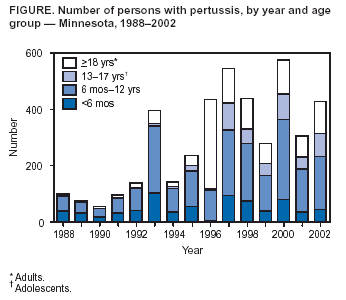 Return to top.
Disclaimer All MMWR HTML versions of articles are electronic conversions from ASCII text into HTML. This conversion may have resulted in character translation or format errors in the HTML version. Users should not rely on this HTML document, but are referred to the electronic PDF version and/or the original MMWR paper copy for the official text, figures, and tables. An original paper copy of this issue can be obtained from the Superintendent of Documents, U.S. Government Printing Office (GPO), Washington, DC 20402-9371; telephone: (202) 512-1800. Contact GPO for current prices. **Questions or messages regarding errors in formatting should be addressed to mmwrq@cdc.gov.Page converted: 2/19/2004 |
|||||||||
This page last reviewed 2/19/2004
|