Diagnosis of Breast Disease. IV. Radiologic Evaluation of the Breast.

[See Algorithm V, "Image-Directed Core Needle Biopsy"]

[See Algorithm VI, "Surgical Evaluation of the Breast"]

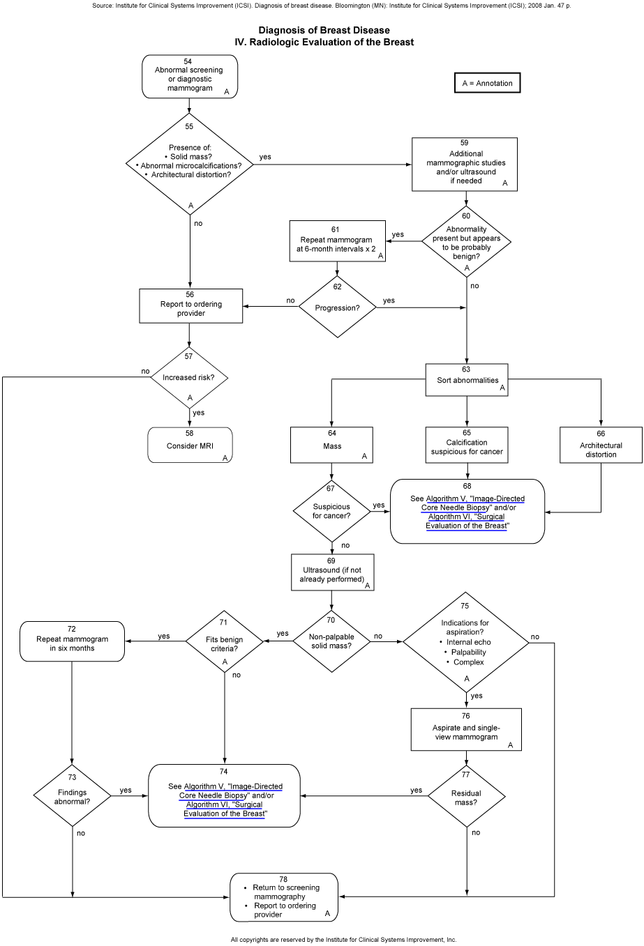
Download a printable copy in PDF format.

Source: Institute for Clinical Systems Improvement (ICSI). Diagnosis of breast disease. Bloomington (MN): Institute for Clinical Systems Improvement (ICSI); 2008 Jan. 47 p. Diagnosis of Breast Disease IV. Radiologic Evaluation of the Breast Abnormal screening or diagnostic mammogram. A A = Annotation Presence of: *Solid mass? *Abnormal microcalcifications? *Architectural distortion? A Additional mammographic studies and/or ultrasound if needed. A Repeat mammogram at 6-month intervals x 2. A Abnormality present but appears to be probably benign? A Report to ordering provider Progression? Increased risk? A Sort abnormalities. A Consider MRI. A Mass. A Calcification suspicious for cancer Architectural distortion Suspicious for cancer? See Algorithm V, Image-Directed  Core Needle Biopsy and/or Algorithm VI, Surgical Evaluation of the Breast Ultrasound (if not already performed). A Repeat mammogram in six months Fits benign criteria? A Non-palpable solid mass? Indications for aspiration? *Internal echo. *Palpability. *Complex. A Aspirate and single-view mammogram Findings abnormal? See Algorithm V, Image-Directed Core Needle Biopsy and/or Algorithm VI, Surgical Evaluation of the Breast Residual mass? *Return to screening mammography. *Report to ordering provider. A All copyrights are reserved by the Institute for Clinical Systems Improvement, Inc.