You are here:
Health Information
Related Information
- What Is Epidermolysis Bullosa? (fast facts, easy-to-read)
- Order a NIAMS Publication to be mailed
Epidermolysis Bullosa
PDF Version of this Document Order this publication
Publication Date: June 2003
Questions and Answers about Epidermolysis Bullosa
This booklet is for people who have epidermolysis bullosa (ep-ee-der-MOL-eh-sis bull-O-sa, often called "EB"), parents and caregivers of children with EB, and others interested in learning more about the disease. The booklet describes the disease and its symptoms and contains information about diagnosis and treatment, as well as current research efforts supported by the National Institute of Arthritis and Musculoskeletal and Skin Diseases (NIAMS) and other components of the Department of Health and Human Services' National Institutes of Health (NIH). It also discusses issues such as skin care and quality of life for people with EB. If you have questions after reading this booklet, you may wish to discuss them with your doctor or a dermatologist (a specialist in treating skin conditions).
What Is Epidermolysis Bullosa?
EB is a group of blistering skin conditions. The skin is so fragile in people with EB that even minor rubbing may cause blistering. At times, the person with EB may not be aware of rubbing or injuring the skin even though blisters develop. In severe EB, blisters are not confined to the outer skin. They may develop inside the body, in such places as the linings of the mouth, esophagus, stomach, intestines, upper airway, bladder, and the genitals.
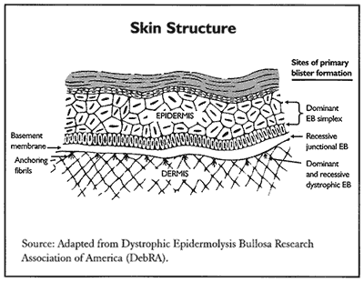
The skin has an outer layer called the epidermis and an underlying layer called the dermis. The place where the two layers meet is called the basement membrane zone. (See the diagram of the skin on page 3.) The main forms of EB are EB Simplex, Junctional EB, and Dystrophic EB. EB Simplex occurs in the outer layer of skin; Junctional EB and Dystrophic EB occur in the basement membrane zone. These major types of EB, which will be described throughout this text, also have many subtypes.
- Who Gets Epidermolysis Bullosa?
- What Causes Epidermolysis Bullosa?
- How Is Epidermolysis Bullosa Diagnosed?
- What Are the Symptoms of Epidermolysis Bullosa?
- How Is Epidermolysis Bullosa Treated?
- What Is the Value of Genetic Counseling?
- What Research Is Being Conducted on Epidermolysis Bullosa?
- What is the Epidermolysis Bullosa Registry and What Does It Do?
- Where Can People Find More Information About Epidermolysis Bullosa?
Who Gets Epidermolysis Bullosa?
It is estimated that 2 to 4 out of every 100,000 people, or up to 12,000 people in the United States, have some form of EB. It occurs in all racial and ethnic groups and affects males and females equally. The disease is not always evident at birth. Milder cases of EB may become apparent when a child crawls, walks, or runs, or when a young adult engages in vigorous physical activity.
What Causes Epidermolysis Bullosa?
Most people with EB have inherited the condition through faulty genes they receive from one or both parents. Genes are located in the body's cells and determine inherited traits passed from parent to child. They also govern every body function, such as the formation of proteins in the skin. More than 10 genes are known to underlie the different forms of EB. Genes are located on chromosomes, which are structures in each cell's nucleus.
In an autosomal dominant form of EB, the disease gene is inherited from only one parent who has the disease, and there is a 50 percent (1 in 2) chance with each pregnancy that a baby will have EB. In the autosomal recessive form, the disease gene is inherited from both parents. Neither parent has to show signs of the disease; they simply need to "carry" the gene, and there is a 25 percent (1 in 4) chance with each pregnancy that a baby will have EB. EB can also be acquired through a mutation (abnormal change) in a gene that occurred during the formation of the egg or sperm reproductive cell in a parent. Neither the sex of the child nor the order of birth determines which child or how many children will develop EB in a family that has the faulty gene.
Although EB Simplex can occur when there is no evidence of the disease in the parents, it is usually inherited as an autosomal dominant disease. In EB Simplex, the faulty genes are those that provide instructions for producing keratin, a fibrous protein in the top layer of skin. As a result, the skin splits in the epidermis, producing a blister.
In Junctional EB, there is a defect in the genes inherited from both parents (autosomal recessive) that normally promote the formation of anchoring filaments (thread-like fibers) or hemidesmosomes [hem-ee-DES-mo-soms] (complex structures composed of many proteins). These structures anchor the epidermis to the underlying basement membrane. The defect leads to tissue separation and blistering in the upper part of the basement membrane.
There are both dominant and recessive forms of Dystrophic EB. In this condition, the filaments that anchor the epidermis to the underlying dermis are either absent or do not function. This is due to defects in the gene for type VII collagen, a fibrous protein that is the main component of the anchoring filaments.
Epidermolysis bullosa acquisita (EBA) is a rare autoimmune disorder where the body attacks its own anchoring fibrils with antibodies, the special proteins that help fight and destroy foreign substances that invade the body. In a few cases, it has occurred following drug therapy for another condition; in most cases, the cause is unknown.
How Is Epidermolysis Bullosa Diagnosed?
Dermatologists can identify where the skin is separating to form blisters and what kind of EB a person has by doing a skin biopsy (taking a small sample of skin that is examined under a microscope). One diagnostic test involves use of a microscope and reflected light to see if proteins needed for forming connecting fibrils, filaments, or hemidesmosomes are missing or reduced in number. Another test involves use of a high-power electron microscope, which can greatly magnify tissue images, to identify structural defects in the skin.
Recent techniques make it possible to identify defective genes in EB patients and their family members. Prenatal diagnosis can now be accomplished by amniocentesis (removing and examining a small amount of amniotic fluid surrounding the fetus in the womb of a pregnant woman) or sampling the chorionic villus (part of the outer membrane surrounding the fetus) as early as the tenth week of pregnancy.
What Are the Symptoms of Epidermolysis Bullosa?
The major sign of all forms of EB is fragile skin that blisters, which can lead to serious complications. For example, blistering areas may become infected, and blisters in the mouth or parts of the gastrointestinal tract may interfere with proper nutrition.
Following is a summary of some of the characteristic signs of various forms of EB.
- EB Simplex (EBS)--A generalized form of EBS usually begins with blistering that is evident at birth or shortly afterward. In a localized, mild form called Weber-Cockayne, blisters rarely extend beyond the feet and hands. In some subtypes of EBS, the blisters occur over widespread areas of the body. Other signs may include thickened skin on the palms of the hands and soles of the feet; rough, thickened, or absent fingernails or toenails; and blistering of the soft tissues inside the mouth. Less common signs include growth retardation; blisters in the esophagus; anemia (a reduction in the red blood cells that carry oxygen to all parts of the body); scarring of the skin; and milia, which are small white skin cysts.
- Junctional EB (JEB)--This disease is usually severe. In the most serious forms, large, ulcerated blisters on the face, trunk, and legs can be life-threatening due to complicated infections and loss of body fluid that leads to severe dehydration. Survival is also threatened by blisters that affect the esophagus, upper airway, stomach, intestines, and the urogenital system. Other signs found in both severe and mild forms of JEB include rough and thickened or absent fingernails and toenails; a thin appearance to the skin (called atrophic scarring); blisters on the scalp or loss of hair with scarring (scarring alopecia); malnutrition and anemia; growth retardation; involvement of soft tissue inside the mouth and nose; and poorly formed tooth enamel.
- Dystrophic EB (DEB)--The dominant and recessive inherited forms of DEB have slightly different symptoms. In some dominant and mild recessive forms, blisters may appear only on the hands, feet, elbows, and knees; nails usually are shaped differently; milia may appear on the skin of the trunk and limbs; and there may be involvement of the soft tissues, especially the esophagus. The more severe recessive form is characterized by blisters over large body surfaces, loss of nails or rough or thick nails, atrophic scarring, milia, itching, anemia, and growth retardation. Severe forms of recessive DEB also may lead to severe eye inflammation with erosion of the cornea (clear covering over the front of the eye), early loss of teeth due to tooth decay, and blistering and scarring inside the mouth and gastrointestinal tract. In most people with this form of EB, some or all the fingers or toes may fuse (pseudosyndactyly). Also, individuals with recessive DEB have a high risk of developing a form of skin cancer called squamous cell carcinoma. It primarily occurs on the hands and feet. The cancer may begin as early as the teenage years. It tends to grow and spread faster in people with EB than in those without the disease.
How Is Epidermolysis Bullosa Treated?
Persons with mild forms of EB may not require extensive treatment. However, they should attempt to keep blisters from forming and prevent infection when blisters occur. Individuals with moderate and severe forms may have many complications and require psychological support along with attention to the care and protection of the skin and soft tissues. Patients, parents, or other care providers should not feel that they must tackle all the complicated aspects of EB care alone. There are doctors, nurses, social workers, clergy members, psychologists, dietitians, and patient and parent support groups that can assist with care and provide information and emotional support.
Preventing Blisters
In many forms of EB, blisters will form with the slightest pressure or friction. This may make parents hesitant to pick up and cuddle young babies. However, a baby needs to feel a gentle human touch and affection, and can be picked up when placed on a soft material and supported under the buttocks (bottom) and behind the neck. A baby with EB should never be picked up under the arms.
A number of things can be done to protect the skin from injury. These include:
- avoiding overheating by keeping rooms at an even temperature
- applying lubricants to the skin to reduce friction and keep the skin moist
- using simple, soft clothing that requires minimal handling when dressing a child
- using sheepskin on car seats and other hard surfaces
- wearing mittens at bedtime to help prevent scratching
Caring for Blistered Skin
When blisters appear, the objectives of care are to reduce pain or discomfort, prevent excessive loss of body fluid, promote healing, and prevent infection.
The doctor may prescribe a mild analgesic to prevent discomfort during changes of dressings (bandages). Dressings that are sticking to the skin may be removed by soaking them off in warm water. While daily cleansing may include a bath with mild soaps, it may be more comfortable to bathe in stages where small areas are cleaned at a time.
Blisters can become quite large and create a large wound when they break. Therefore, a medical professional will likely provide instructions on how to safely break a blister in its early stages while still leaving the top skin intact to cover the underlying reddened area. One technique is to pat the blister with an alcohol pad before popping it at the sides with a sterile needle or other sterile tool. The fluid can then drain into a sterile gauze that is used to dab the blister. After opening and draining, the doctor may suggest that an antibiotic ointment be applied to the area of the blister before covering it with a sterile, nonsticking bandage. To prevent irritation of the skin from tape, a bandage can be secured with a strip of gauze that is tied around it. In milder cases of EB or where areas are difficult to keep covered, the doctor may recommend leaving a punctured blister open to the air.
A moderately moist environment promotes healing, but heavy drainage from blister areas may further irritate the skin, and an absorbent or foam dressing may be needed. There are also contact layer dressings where a mesh layer through which drainage can pass is placed on the wound and is topped by an outer absorbent layer. The doctor or other health care professional may recommend gauze or bandages that are soaked with petroleum jelly, glycerin, or moisturizing substances, or may suggest more extensive wound care bandages or products.
Treating Infection
The chances of skin infection can be reduced by good nutrition, which builds the body's defenses and promotes healing, and by careful skin care with clean hands and use of sterile materials. For added protection, the doctor may recommend antibiotic ointments and soaks.
Even in the presence of good care, it is possible for infection to develop. Signs of infection are redness and heat around an open area of skin, pus or a yellow drainage, excessive crusting on the wound surface, a red line or streak under the skin that spreads away from the blistered area, a wound that does not heal, and/or fever or chills. The doctor may prescribe a specific soaking solution, an antibiotic ointment, or an oral antibiotic to reduce the growth of bacteria. Wounds that are not healing may be treated by a special wound covering or biologically developed skin.
Treating Nutritional Problems
Blisters that form in the mouth and esophagus in some people with EB are likely to cause difficulty in chewing and swallowing food and drinks. If breast or bottle feeding results in blisters, infants may be fed using a preemie nipple (a soft nipple with large holes), a cleft palate nipple, an eyedropper, or a syringe. When the baby is old enough to take in food, adding extra liquid to pureed (finely mashed) food makes it easier to swallow. Soups, milk drinks, mashed potatoes, custards, and puddings can be given to young children. However, food should never be served too hot.
Dietitians are important members of the health care team that assists people with EB. They can work with family members and older patients to find recipes and prepare food that is nutritious and easy to consume. For example, they can identify high-caloric and protein-fortified foods and beverages that help replace protein lost in the fluid from draining blisters. They can suggest vitamin and mineral nutritional supplements that may be needed, and show how to mix these into the food and drinks of young children. Dietitians can also recommend adjustments in the diet to prevent gastrointestinal problems, such as constipation, diarrhea, or painful elimination.
Surgical Treatment
Surgical treatment may be necessary in some forms of EB. Individuals with the severe forms of autosomal recessive Dystrophic EB whose esophagus has been narrowed by scarring may require dilation of their esophagus for food to travel from the mouth to the stomach. Other individuals who are not getting proper nutrition may need a feeding tube that permits delivery of food directly to the stomach. Also, patients whose fingers or toes are fused together may require surgery to release them.
What Is the Value of Genetic Counseling?
Epidermolysis bullosa is a difficult, sometimes painful, and often disfiguring disease. Most adults with signs of EB or who know they carry the gene would like to spare future generations, including their own potential offspring, from this condition. With the knowledge of specific gene mutations that cause EB, it is now possible to determine the specific gene mutation in the family and then to conduct prenatal tests on pregnant women with a fetus at risk of EB to determine the status of the fetus. Specific laboratories, such as the one affiliated with the Dystrophic Epidermolysis Bullosa Research Association (DebRA), can test for gene mutations. (See resource list at the back of this booklet.) Also, a genetic counselor can provide information on the likelihood of passing the gene for EB to children and provide advice on future childbearing. Genetic counseling can be a crucial step in helping families make decisions about their family planning.
What Research Is Being Conducted on Epidermolysis Bullosa?
At one time, research on EB was limited to describing the disease and understanding what happens in the layers of skin. Today, research focuses upon finding gene mutations and their effect on the tissues, copying genes, reproducing gene mutations for research to correct them, inserting healthy genes to replace missing or mutated genes, and screening those who may have a gene mutation causing EB.
Some researchers are aiming their sights on future gene therapy. They are developing mouse models to detect the involvement of different tissues in EB and to test the delivery of modified cells to genetically altered mice that have EB traits. While scientists have already been able to achieve genetic correction of some human genetic skin diseases, they have not been able to sustain the results beyond a few weeks or months. Therefore, they are working to achieve long-lasting corrective gene delivery that can be used as a springboard for further gene therapy trials in humans.
In Dystrophic EB, the fibrils that anchor the epidermis to the underlying dermis are either absent or do not function well. Scientists are introducing a gene for absent collagen (type VII) into cultured keratinocytes and fibroblasts (types of skin cells) obtained from patients whose cells cannot make the protein. It is hoped that the gene-corrected cells eventually can be transplanted back into the patients to promote and sustain the formation of anchoring fibrils.
Applying newer diagnostic techniques, investigators are beginning to link specific gene defects with the protein problems they produce. Now that the EB-causing gene mutations can be identified, there is a way that ova (eggs) that do not contain an abnormal gene can be selected for in vitro fertilization outside the body, thus improving the chances of having healthy children in families with the EB gene.
Researchers are also assessing the effectiveness of using proteins called cytokines and new kinds of dressings to heal blister wounds.
What Is the Epidermolysis Bullosa Registry and What Does It Do?
The National EB Registry collects information from patients with EB, characterizes the many different forms of EB and determines risks of various symptoms associated with the disease. The information is used for research to improve understanding and provide better treatment of EB. The registry is also a resource for initial diagnostic testing of patients.
-
National Epidermolysis Bullosa Registry
c/o Vanderbilt University Medical CenterNashville, TN
Registry contacts: Jo-David Fine, M.D, M.P.H., and Madeline Weiner, R.N.
Phone Numbers: Dr. Fine 615-329-0801; Madeline Weiner 919-929-1855
Email: Jo-David.Fine@vanderbilt.edu; debra.nurse@mindspring.com
Where Can People Find More Information About Epidermolysis Bullosa?
-
National Institute of Arthritis and Musculoskeletal and Skin Diseases (NIAMS)
Information Clearinghouse
National Institutes of Health1 AMS Circle
Bethesda, MD 20892-3675
Phone: 301-495-4484
Toll Free: 877-22-NIAMS (226-4267)
TTY: 301–565–2966
Fax: 301-718-6366
Email: NIAMSinfo@mail.nih.gov
Website: http://www.niams.nih.govNIAMS provides information about various forms of arthritis and rheumatic diseases and bone, muscle, joint, and skin diseases. It distributes patient and professional education materials and also refers people to other sources of information. Additional information and updates can be found on the NIAMS Web site.
-
Dystrophic Epidermolysis Bullosa Research Association of America, Inc. (DebRA)
5 West 36th Street, Suite 404
New York, NY 10018
Phone: 212-868-1573
Fax: 212-868-9296
Email: staff@debra.org
Website: http://www.debra.orgDebRA is a national, nonprofit, voluntary organization formed as an information source for patients, family members, the general public, and health professionals. It also promotes and supports research and can provide assistance in locating testing sites, medical treatment, support groups, and genetic counseling. A full-time EB nurse educator is available to provide assistance and support to EB patients, caregivers, and medical professionals.
A molecular diagnostic laboratory affiliated with DebRA is located at Thomas Jefferson University in Philadelphia, Pennsylvania. Your doctor can make arrangements to submit blood samples for genetic analysis. A small fetal sample can be sent from a doctor's office to the laboratory to find out if a fetus is affected by EB.
-
DebRA Molecular Diagnostic Laboratory at Thomas Jefferson University
833 Chestnut Street, Room 428
Philadelphia, PA 19107
Phone: 215-503-2176
Fax: 215-503-5788
-
National Information Center for Children and Youth with Disabilities
P.O. Box 1492
Washington, DC 20013
Phone: 202-884-8200
Fax: 202-884-8441
Email: NICHCY@aed.org
Website: http://www.NICHCY.orgThe Center distributes information about services, special education programs, and laws affecting disabled children and adults. It is supported by the U.S. Department of Education.
-
National Society of Genetic Counselors, Inc.
233 Canterbury Drive
Wallingford, PA 19086-6617
Phone: 610-872-7608
Email: nsgc@aol.com
Website: http://www.nsgc.orgThis organization, which represents professional genetic counselors, provides assistance in locating counselors and provides a list of questions that might be asked when meeting with a counselor.
Acknowledgments
The NIAMS gratefully acknowledges the assistance of Jo-David Fine, M.D., M.P.H., National EB Registry, Lexington, Kentucky; Martin I. Hassner, DebRA; Alan N. Moshell, M.D., NIAMS, NIH; Amy S. Paller, M.D., Children's Memorial Hospital, Chicago, Illinois; Jouni J. Uitto, M.D., Ph.D., Thomas Jefferson University, Philadelphia, Pennsylvania; and David T. Woodley, M.D., Los Angeles County/University of Southern California Medical Center, Los Angeles, California, in the preparation of this booklet.
The mission of the National Institute of Arthritis and Musculoskeletal and Skin Diseases (NIAMS), a part of the Department of Health and Human Services' National Institutes of Health (NIH), is to support research into the causes, treatment, and prevention of arthritis and musculoskeletal and skin diseases, the training of basic and clinical scientists to carry out this research, and the dissemination of information on research progress in these diseases. The National Institute of Arthritis and Musculoskeletal and Skin Diseases Information Clearinghouse is a public service sponsored by the NIAMS that provides health information and information sources. Additional information can be found on the NIAMS Web site at www.niams.nih.gov.
NIH Publication No. 03-7038