MRI of the Body (Chest, Abdomen, Pelvis)
What is MRI of the Body?
Magnetic resonance imaging (MRI) is a noninvasive medical test that helps physicians diagnose and treat medical conditions.
MR imaging uses a powerful magnetic field, radio frequency pulses and a computer to produce detailed pictures of organs, soft tissues, bone and virtually all other internal body structures. The images can then be examined on a computer monitor, printed or copied to CD. MRI does not use ionizing radiation (x-rays).
Detailed MR images allow physicians to better evaluate parts of the body and certain diseases that may not be assessed adequately with other imaging methods such as x-ray, ultrasound or computed tomography (also called CT or CAT scanning).
What are some common uses of the procedure?
MR imaging of the body is performed to evaluate:
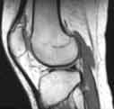
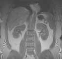
- organs of the chest, abdomen and pelvis—including the heart, liver, biliary tract, kidney, spleen, and pancreas and adrenal glands.
- pelvic organs including the reproductive organs in the male (prostate and testicles) and the female (uterus, cervix and ovaries).
- pelvic and hip bones.
- blood vessels (MR Angiography).
- breasts.
Physicians use the MR examination to help diagnose or monitor treatment for conditions such as:
- tumors of the chest, abdomen or pelvis.
- coronary artery disease and heart problems including the aorta, coronary arteries and blood vessels, by examining the size and thickness of the chambers of the heart and the extent of damage caused by a heart attack or progressive heart disease. For more information, visit the MR Angiography and Cardiac CT for Calcium Scoring pages.
- tumors and other abnormalities of the reproductive organs (e.g., uterus, ovaries, testicles, prostate).
- causes of pelvic pain in women, such as endometriosis.
- functional and anatomical abnormalities of the heart.
- diseases of the liver, such as cirrhosis, and that of other abdominal organs (when a complete diagnostic assessment can not be done with other techniques).
- congenital arterial and venous vascular anomalies and diseases (e.g., atherosclerosis) of the chest, abdomen and pelvis (MR Angiography).
- conditions involving the bile duct, gallbladder and pancreatic ducts (MRCP).
- breast cancer and implants.
How should I prepare for the procedure?
You may be asked to wear a gown during the exam or you may be allowed to wear your own clothing if it is loose-fitting and has no metal fasteners.
Guidelines about eating and drinking before an MRI exam vary with the specific exam and also with the facility. Unless you are told otherwise, you may follow your regular daily routine and take medications as usual.
Some MRI examinations may require the patient to swallow contrast material or receive an injection of contrast into the bloodstream. The radiologist or technologist may ask if you have allergies of any kind, such as allergy to iodine or x-ray contrast material, drugs, food, the environment, or asthma. However, the contrast material used for an MRI exam, called gadolinium, does not contain iodine and is less likely to cause an allergic reaction.
The radiologist should also know if you have any serious health problems and what surgeries you have undergone. Some conditions, such as severe kidney disease may prevent you from having an MRI with contrast material.
Women should always inform their physician or technologist if there is any possibility that they are pregnant. MRI has been used for scanning patients since the 1980's with no reports of any ill effects on pregnant women or their babies. However, because the baby will be in a strong magnetic field, pregnant women should not have this exam unless the potential benefit from the MRI is assumed to outweigh the potential risks. See the Safety page for more information about pregnancy and MR imaging.
If you have claustrophobia (fear of enclosed spaces) or anxiety, you may want to ask your physician for a prescription for a mild sedative.
Jewelry and other accessories should be left at home if possible, or removed prior to the MRI scan. Because they can interfere with the magnetic field of the MRI unit, metal and electronic objects are not allowed in the exam room. These items include:
- jewelry, watches, credit cards and hearing aids, all of which can be damaged.
- pins, hairpins, metal zippers and similar metallic items, which can distort MRI images.
- removable dental work.
- pens, pocketknives and eyeglasses.
- body piercings.
In most cases, an MRI exam is safe for patients with metal implants, except for a few types. People with the following implants cannot be scanned and should not enter the MRI scanning area unless explicitly instructed to do so by a radiologist or technologist who is aware of the presence of any of the following:
- internal (implanted) defibrillator or pacemaker
- cochlear (ear) implant
- some types of clips used on brain aneurysms
You should tell the technologist if you have medical or electronic devices in your body, because they may interfere with the exam or potentially pose a risk. Examples include but are not limited to:
- artificial heart valves
- implanted drug infusion ports
- implanted electronic device, including a cardiac pacemaker
- artificial limbs or metallic joint prostheses
- implanted nerve stimulators
- metal pins, screws, plates or surgical staples.
In general, metal objects used in orthopedic surgery pose no risk during MRI. However, a recently placed artificial joint may require the use of another imaging procedure. If there is any question of their presence, an x-ray may be taken to detect the presence of any metal objects.
Patients who might have metal objects in certain parts of their bodies may also require an x-ray prior to an MRI. Dyes used in tattoos may contain iron and could heat up during MRI, but this is rarely a problem. Tooth fillings and braces usually are not affected by the magnetic field but they may distort images of the facial area or brain, so the radiologist should be aware of them.
What does the equipment look like?
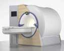
The traditional MRI unit is a large cylinder-shaped tube surrounded by a circular magnet. You will lie on a moveable examination table that slides into the center of the magnet.
Some MRI units, called short-bore systems, are designed so that the magnet does not completely surround you; others are open on all sides (open MRI). These units are especially helpful for examining patients who are fearful of being in a closed space and for those who are very obese. Newer open MRI units provide very high quality images for many types of exams; however, open MRI units with older magnets may not provide this same quality. Certain types of exams cannot be performed using open MRI. For more information, consult your doctor.
The computer workstation that processes the imaging information is located in a separate room than the scanner.
How does the procedure work?
Unlike conventional x-ray examinations and computed tomography (CT) scans, MRI does not depend on radiation. Instead, while in the magnet, radio waves redirect the axes of spinning protons, which are the nuclei of hydrogen atoms, in a strong magnetic field.
The magnetic field is produced by passing an electric current through wire coils in most MRI units. Other coils, located in the machine and in some cases, placed around the part of the body being imaged, send and receive radio waves, producing signals that are detected by the coils.
A computer then processes the signals and generates a series of images each of which shows a thin slice of the body. The images can then be studied from different angles by the interpreting physician.
Overall, the differentiation of abnormal (diseased) tissue from normal tissues is often easier with MRI than with other imaging modalities such as x-ray, CT and ultrasound.
What will I experience during and after the procedure?
Most MRI exams are painless.
Some patients, however, find it uncomfortable to remain still during MR imaging. Others experience a sense of being closed-in (claustrophobia). Therefore, sedation can be arranged for those patients who anticipate anxiety, but fewer than one in 20 require it.
It is normal for the area of your body being imaged to feel slightly warm, but if it bothers you, notify the radiologist or technologist. It is important that you remain perfectly still while the images are being recorded, which is typically only a few seconds to a few minutes at a time. For some types of exams, you may be asked to hold your breath. You will know when images are being recorded because you will hear tapping or thumping sounds when the coils that generate the radiofrequency pulses are activated. You will be able to relax between imaging sequences, but will be asked to maintain your position as much as possible.
You will be alone in the exam room during the MR imaging, however, the technologist will be able to see, hear and speak with you at all times using a two-way intercom. Many MRI centers allow a friend or parent to stay in the room.
You may be offered or you may request earplugs to reduce the noise of the MRI scanner, which produces loud thumping and humming noises during imaging. MRI scanners are air-conditioned and well-lit. Some scanners have music to help you pass the time.
When the contrast material is injected, it is normal to feel coolness and a flushing for a minute or two. The intravenous needle may cause you some discomfort when it is inserted and once it is removed, you may experience some bruising. There is also a very small chance of irritation of your skin at the site of the IV tube insertion.
If you have not been sedated, no recovery period is necessary. You may resume your usual activities and normal diet immediately after the exam. A few patients experience side effects from the contrast material, including nausea and local pain. Very rarely, patients are allergic to the contrast material and experience hives, itchy eyes or other reactions.
It is recommended that nursing mothers not breastfeed for 36 to 48 hours after an MRI with a contrast material.
Who interprets the results and how do I get them?
A radiologist, a physician specifically trained to supervise and interpret radiology examinations, will analyze the images and send a signed report to your primary care or referring physician, who will share the results with you.
What are the benefits vs. risks?
Benefits
- MRI is a noninvasive imaging technique that does not involve exposure to radiation.
- MR images of the soft-tissue structures of the body—such as the heart, liver and many other organs— is more likely to identify and characterize abnormalities and focal lesions than other imaging methods. This detail makes MRI an invaluable tool in early diagnosis and evaluation of many focal lesions and tumors.
- MRI has proven valuable in diagnosing a broad range of conditions, including cancer, heart and vascular disease, and muscular and bone abnormalities.
- MRI enables the detection of abnormalities that might be obscured by bone with other imaging methods.
- MRI allows physicians to assess the biliary system noninvasively and without contrast injection.
- The contrast material used in MRI exams is less likely to produce an allergic reaction than the iodine-based materials used for conventional x-rays and CT scanning.
- MRI provides a fast, noninvasive alternative to x-ray angiography for diagnosing problems of the heart and blood vessels.
Risks
- The MRI examination poses almost no risk to the average patient when appropriate safety guidelines are followed.
- If sedation is used there are risks of excessive sedation. The technologist or nurse monitors your vital signs to minimize this risk.
- Although the strong magnetic field is not harmful in itself, medical devices that contain metal may malfunction or cause problems during an MRI exam.
- There is a very slight risk of an allergic reaction if contrast material is injected. Such reactions usually are mild and easily controlled by medication.
- Nephrogenic systemic fibrosis is currently a recognized, but rare, complication of MRI believed to be caused by the injection of high doses of MRI contrast material in patients with poor kidney function.
What are the limitations of MRI of the Body?
High-quality images are assured only if you are able to remain perfectly still while the images are being recorded. If you are anxious, confused or in severe pain, you may find it difficult to lie still during imaging.
A person who is very large may not fit into the opening of a conventional MRI machine.
The presence of an implant or other metallic object often makes it difficult to obtain clear images and patient movement can have the same effect.
Breathing may cause artifacts, or image distortions, during MRIs of the chest, abdomen and pelvis. Bowel motion is another source of motion artifacts in abdomen and pelvic MRI studies.
Although there is no reason to believe that magnetic resonance imaging harms the fetus, pregnant women usually are advised not to have an MRI exam unless medically necessary.
MRI may not always distinguish between cancer tissue and edema fluid.
MRI typically costs more and may take more time to perform than other imaging modalities.
|