You are here:
Health Information
Related Information
- What are Shoulder Problems (fast facts, easy-to-read)
- Joint Replacement
Other Information
Shoulder Problems
PDF Version of this Document Order this publication
Publication Date: May 2001
Revised March 2006
Questions and Answers about Shoulder Problems
This booklet first answers general questions about the shoulder and shoulder problems. It then answers questions about specific shoulder problems as well as shoulder pain caused by arthritis of the shoulder.
What Are the Most Common Shoulder Problems?
The most movable joint in the body, the shoulder is also one of the most potentially unstable joints. As a result, it is the site of many common problems. They include sprains, strains, dislocations, separations, tendinitis, bursitis, torn rotator cuffs, frozen shoulder, fractures, and arthritis. Specific shoulder problems will be discussed later in this booklet.
- How Common Are Shoulder Problems?
- What Are the Structures of the Shoulder and How Does It Function?
- What Are the Origins and Causes of Shoulder Problems?
- How Are Shoulder Problems Diagnosed?
- What Should I Know About Specific Shoulder Problems, Including Their Symptoms and Treatment?
- What Research Is Being Done on Shoulder Problems?
- Where Can People Get Additional Information About Shoulder Problems?
- Key Words
Information Boxes
How Common Are Shoulder Problems?
According to the Centers for Disease Control and Prevention, about 13.7 million people in the United States sought medical care in 2003 for shoulder problems.
What Are the Structures of the Shoulder and How Does It Function?
To better understand shoulder problems and how they occur, it helps to begin with an explanation of the shoulder's structure and how it functions.
The shoulder joint is composed of three bones: the clavicle (collarbone), the scapula (shoulder blade), and the humerus (upper arm bone). (See diagram.) Two joints facilitate shoulder movement. The acromioclavicular (ah-KRO-me-o-klah-VIK-u-lahr; AC) joint is located between the acromion (ah-KRO-me-on; part of the scapula that forms the highest point of the shoulder) and the clavicle. The glenohumeral joint, commonly called the shoulder joint, is a ball-and-socket-type joint that helps move the shoulder forward and backward and allows the arm to rotate in a circular fashion or hinge out and up away from the body. (The "ball," or humerus, is the top, rounded portion of the upper arm bone; the "socket," or glenoid, is a dish-shaped part of the outer edge of the scapula into which the ball fits.) The capsule is a soft tissue envelope that encircles the glenohumeral joint. It is lined by a thin, smooth synovial membrane.
Shoulder
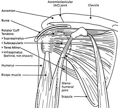
In contrast to the hip joint, which more closely approximates a true ball and socket joint, the shoulder joint can be compared to a golf ball and tee, in which the ball can easily slip off the flat tee. Because the bones provide little inherent stability to the shoulder joint, it is highly dependent on surrounding soft tissues such as capsule ligaments and the muscles surrounding the rotator cuff to hold the ball in place. Whereas the hip joint is inherently quite stable because of the encircling bony anatomy, it also is relatively immobile. The shoulder, on the other hand, is relatively unstable but highly mobile, allowing an individual to place the hand in numerous positions. It is in fact, one of the most mobile joints in the human body.
The bones of the shoulder are held in place by muscles, tendons, and ligaments. Tendons are tough cords of tissue that attach the shoulder muscles to bone and assist the muscles in moving the shoulder. Ligaments attach shoulder bones to each other, providing stability. For example, the front of the joint capsule is anchored by three glenohumeral ligaments. The rotator cuff is a structure composed of tendons that work along with associated muscles to hold the ball at the top of the humerus in the glenoid socket and provide mobility and strength to the shoulder joint. Two filmy sac-like structures called bursae permit smooth gliding between bones, muscles, and tendons. They cushion and protect the rotator cuff from the bony arch of the acromion.
What Are the Origins and Causes of Shoulder Problems?
The shoulder is easily injured because the ball of the upper arm is larger than the shoulder socket that holds it. To remain stable, the shoulder must be anchored by its muscles, tendons, and ligaments.
Although the shoulder is easily injured during sporting activities and manual labor, the primary source of shoulder problems appears to be the natural age-related degeneration of the surrounding soft tissues such as those found in the rotator cuff. The incidence of rotator cuff problems rises dramatically as a function of age and is generally seen among individuals who are more than 60 years old. Often, the dominant and nondominant arm will be affected to a similar degree. Overuse of the shoulder can lead to more rapid age-related deterioration.
Shoulder pain may be localized or may be felt in areas around the shoulder or down the arm. Disease within the body (such as gallbladder, liver, or heart disease, or disease of the cervical spine of the neck) also may generate pain that travels along nerves to the shoulder. However, these other causes of shoulder pain are beyond the scope of this book, which will focus on problems within the shoulder itself.
How Are Shoulder Problems Diagnosed?
As with any medical issue, a shoulder problem is generally diagnosed using a three-part process:
- medical history - The patient tells the doctor about any injury or other condition that might be causing the pain.
- physical examination - The doctor examines the patient to feel for injury and to discover the limits of movement, location of pain, and extent of joint instability.
- tests - The doctor may order one or more of the tests listed below to make a specific diagnosis. These tests may include the following:
- Standard x ray - a familiar procedure in which low-level radiation is passed through the body to produce a picture called a radiograph. An x ray is useful for diagnosing fractures or other problems of the bones. Soft tissues, such as muscles and tendons, do not show up on x rays.
- Arthrogram - a diagnostic record that can be seen on an x ray after injection of a contrast fluid into the shoulder joint to outline structures such as the rotator cuff. In disease or injury, this contrast fluid may either leak into an area where it does not belong, indicating a tear or opening, or be blocked from entering an area where there normally is an opening.
- Ultrasound - a noninvasive, patient-friendly procedure in which a small, hand-held scanner is placed on the skin of the shoulder. Just as ultrasound waves can be used to visualize the fetus during pregnancy, they can also be reflected off the rotator cuff and other structures to form a high-quality image of them. The accuracy of ultrasound for the rotator cuff is particularly high.
- MRI (magnetic resonance imaging) - a noninvasive procedure in which a machine with a strong magnet passes a force through the body to produce a series of cross-sectional images of the shoulder. Other diagnostic tests, such as one that involves injecting an anesthetic into and around the shoulder joint, are discussed in detail in other parts of this booklet.
Other diagnostic tests, such as one that involves injecting an anesthetic into and around the shoulder joint, are discussed in detail in other parts of this booklet.
What Should I Know About Specific Shoulder Problems, Including Their Symptoms and Treatment?
The symptoms of shoulder problems, as well as their diagnosis and treatment, vary widely, depending on the specific problem. The following is important information to know about some of the most common shoulder problems.
Dislocation
The shoulder joint is the most frequently dislocated major joint of the body. In a typical case of a dislocated shoulder, either a strong force pulls the shoulder outward (abduction) or extreme rotation of the joint pops the ball of the humerus out of the shoulder socket. Dislocation commonly occurs when there is a backward pull on the arm that either catches the muscles unprepared to resist or overwhelms the muscles. When a shoulder dislocates frequently, the condition is referred to as shoulder instability. A partial dislocation in which the upper arm bone is partially in and partially out of the socket is called a subluxation.
-
Signs and symptoms: The shoulder can dislocate either forward, backward, or downward. When the shoulder dislocates, the arm appears out of position. Other symptoms include pain, which may be worsened by muscle spasms; swelling; numbness; weakness; and bruising. Problems seen with a dislocated shoulder are tearing of the ligaments or tendons reinforcing the joint capsule and, less commonly, bone and/or nerve damage.
Diagnosis: Doctors usually diagnose a dislocation by a physical examination; x rays may be taken to confirm the diagnosis and to rule out a related fracture.
Treatment: Doctors treat a dislocation by putting the ball of the humerus back into the joint socket, a procedure called a reduction. The arm is then stabilized for several weeks in a sling or a device called a shoulder immobilizer. Usually the doctor recommends resting the shoulder and applying ice three or four times a day. After pain and swelling have been controlled, the patient enters a rehabilitation program that includes exercises. The goal is to restore the range of motion of the shoulder, strengthen the muscles, and prevent future dislocations. These exercises may progress from simple motion to the use of weights.
After treatment and recovery, a previously dislocated shoulder may remain more susceptible to re-injury, especially in young, active individuals. Ligaments may have been stretched or torn, and the shoulder may tend to dislocate again. A shoulder that dislocates severely or often, injuring surrounding tissues or nerves, usually requires surgical repair to tighten stretched ligaments or reattach torn ones.
Sometimes the doctor performs surgery through a tiny incision into which a small scope (arthroscope) is inserted to observe the inside of the joint. After this procedure, called arthroscopic surgery, the shoulder is generally stabilized for about 6 weeks. Full recovery takes several months. Arthroscopic techniques involving the shoulder are relatively new, and some surgeons prefer to repair a recurrent dislocating shoulder by time-tested open surgery under direct vision. Usually following open surgery there are fewer repeat dislocations, and movement is improved, but there is often some loss of motion.
Separation
A shoulder separation occurs where the collarbone (clavicle) meets the shoulder blade (scapula). When ligaments that hold the joint together are partially or completely torn, the outer end of the clavicle may slip out of place, preventing it from properly meeting the scapula. Most often, the injury is caused by a blow to the shoulder or by falling on an outstretched hand.
-
Signs and symptoms: Shoulder pain or tenderness and, occasionally, a bump in the middle of the top of the shoulder (over the acromioclavicular (AC) joint) are signs that a separation may have occurred.
Diagnosis: Doctors may diagnose a separation by performing a physical examination. They may confirm the diagnosis and determine the severity of the separation by taking an x ray. While the x ray is being taken, the patient makes the separation more pronounced by holding a light weight that pulls on the muscles.
Treatment: A shoulder separation is usually treated conservatively by rest and wearing a sling. Soon after injury, an ice bag may be applied to relieve pain and swelling. After a period of rest, a therapist helps the patient perform exercises that put the shoulder through its range of motion. Most shoulder separations heal within 2 or 3 months without further intervention. However, if ligaments are severely torn, surgical repair may be required to hold the clavicle in place. A doctor may wait to see if conservative treatment works before deciding whether surgery is required.
Rotator Cuff Disease: Tendinitis and Bursitis
These conditions are closely related and may occur alone or in combination.
Tendinitis is inflammation (redness, soreness, and swelling) of a tendon. In tendinitis of the shoulder, the rotator cuff and/or biceps tendon become inflamed, usually as a result of being pinched by surrounding structures. The injury may vary from mild inflammation to involvement of most of the rotator cuff. When the rotator cuff tendon becomes inflamed and thickened, it may get trapped under the acromion. Squeezing of the rotator cuff is called impingement syndrome.
Bursitis, or inflammation of the bursa sacs that protect the shoulder, may accompany tendinitis and impingement syndrome. Inflammation caused by a disease such as rheumatoid arthritis may cause rotator cuff tendinitis and bursitis. Sports involving overuse of the shoulder and occupations requiring frequent overhead reaching are other potential causes of irritation to the rotator cuff or bursa and may lead to inflammation and impingement.
If the rotator cuff and bursa are irritated, inflamed, and swollen, they may become squeezed between the head of the humerus and the acromion. Repeated motion involving the arms, or the effects of the aging process on shoulder movement over many years, may also irritate and wear down the tendons, muscles, and surrounding structures.
-
Signs and Symptoms: Signs of these conditions include the slow onset of discomfort and pain in the upper shoulder or upper third of the arm and/or difficulty sleeping on the shoulder. Tendinitis and bursitis also cause pain when the arm is lifted away from the body or overhead. If tendinitis involves the biceps tendon (the tendon located in front of the shoulder that helps bend the elbow and turn the forearm), pain will occur in the front or side of the shoulder and may travel down to the elbow and forearm. Pain may also occur when the arm is forcefully pushed upward overhead.
Diagnosis: Diagnosis of tendinitis and bursitis begins with a medical history and physical examination. X rays do not show tendons or the bursae, but may be helpful in ruling out bony abnormalities or arthritis. The doctor may remove and test fluid from the inflamed area to rule out infection. Impingement syndrome may be confirmed when injection of a small amount of anesthetic (lidocaine hydrochloride) into the space under the acromion relieves pain.
Treatment: The first step in treating these conditions is to reduce pain and inflammation with rest, ice, and anti-inflammatory medicines such as aspirin and ibuprofen (Advil*, Motrin). In some cases, the doctor or therapist will use ultrasound (gentle sound-wave vibrations) to warm deep tissues and improve blood flow. Gentle stretching and strengthening exercises are added gradually. These may be preceded or followed by use of an ice pack. If there is no improvement, the doctor may inject a corticosteroid medicine into the space under the acromion. While steroid injections are a common treatment, they must be used with caution because they may lead to tendon rupture. If there is still no improvement after 6 to 12 months, the doctor may recommend either arthroscopic or open surgery to repair damage and relieve pressure on the tendons and bursae.
* Brand names included in this booklet are provided as examples only, and their inclusion does not mean that these products are endorsed by the National Institutes of Health or any other Government agency. Also, if a particular brand name is not mentioned, this does not mean or imply that the product is unsatisfactory.
Torn Rotator Cuff
Rotator cuff tendons often become inflamed from overuse, aging, or a fall on an outstretched hand or another traumatic cause. Sports or occupations requiring repetitive overhead motion or heavy lifting can also place a significant strain on rotator cuff muscles and tendons. Over time, as a function of aging, tendons become weaker and degenerate. Eventually, this degeneration can lead to complete tears of both muscles and tendons. These tears are surprisingly common. In fact, a tear of the rotator cuff is not necessarily an abnormal situation in older individuals if there is no significant pain or disability. Fortunately, these tears do not lead to any pain or disability in most people. However, some individuals can develop very significant pain as a result of these tears and they may require treatment.
-
Signs and Symptoms: Typically, a person with a rotator cuff injury feels pain over the deltoid muscle at the top and outer side of the shoulder, especially when the arm is raised or extended out from the side of the body. Motions like those involved in getting dressed can be painful. The shoulder may feel weak, especially when trying to lift the arm into a horizontal position. A person may also feel or hear a click or pop when the shoulder is moved. Pain or weakness on outward or inward rotation of the arm may indicate a tear in a rotator cuff tendon. The patient also feels pain when lowering the arm to the side after the shoulder is moved backward and the arm is raised.
Diagnosis: A doctor may detect weakness but may not be able to determine from a physical examination where the tear is located. X rays, if taken, may appear normal. An MRI or ultrasound can help detect a full tendon tear or a partial tendon tear.
Treatment: Doctors usually recommend that patients with a rotator cuff injury rest the shoulder, apply heat or cold to the sore area, and take medicine to relieve pain and inflammation. Other treatments might be added, such as electrical stimulation of muscles and nerves, ultrasound, or a cortisone injection near the inflamed area of the rotator cuff. If surgery is not an immediate consideration, exercises are added to the treatment program to build flexibility and strength and restore the shoulder's function. If there is no improvement with these conservative treatments and functional impairment persists, the doctor may perform arthroscopic or open surgical repair of the torn rotator cuff.
Treatment for rotator cuff disease usually depends on the severity of the injury, the age and health status of the patient, and the length of time a given patient may have had the condition. Patients with rotator cuff tendinitis or bursitis that does not include a complete tear of the tendon can usually be treated without surgery. Nonsurgical treatments include the use of anti-inflammatory medication and occasional steroid injections into the area of the inflamed rotator cuff, followed by rehabilitative rotator cuff strengthening exercises. These treatments are best undertaken with the guidance of a health-care professional such as a physical therapist, who works in conjunction with the treating physician.
Surgical repair of rotator cuff tears is best for:
- younger patients, especially those with small tears. Surgery leads to a high degree of successful healing and reduces concerns about the tear getting worse over time.
- individuals whose rotator cuff tears are caused by an acute, severe injury. These people should seek immediate treatment that includes surgical repair of the tendon.
Generally speaking, individuals who are older and have had shoulder pain for a longer period of time can be treated with nonoperative measures even in the presence of a complete rotator cuff tear. These people are often treated similarly to those who have pain, but do not have a rotator cuff tear. Again, anti-inflammatory medication, use of steroid injections, and rehabilitative exercises can be very effective. When treated surgically, rotator cuff tears can be repaired by either arthroscopic or traditional open surgical techniques.
Frozen Shoulder (Adhesive Capsulitis)
As the name implies, movement of the shoulder is severely restricted in people with a "frozen shoulder." This condition, which doctors call adhesive capsulitis, is frequently caused by injury that leads to lack of use due to pain. Rheumatic disease progression and recent shoulder surgery can also cause frozen shoulder. Intermittent periods of use may cause inflammation. Adhesions (abnormal bands of tissue) grow between the joint surfaces, restricting motion. There is also a lack of synovial fluid, which normally lubricates the gap between the arm bone and socket to help the shoulder joint move. It is this restricted space between the capsule and ball of the humerus that distinguishes adhesive capsulitis from a less complicated painful, stiff shoulder. People with diabetes, stroke, lung disease, rheumatoid arthritis, and heart disease, or those who have been in an accident, are at a higher risk for frozen shoulder. Frozen shoulder is more common among women than men. People between the ages of 40 and 70 are most likely to experience it.
-
Signs and symptoms: With a frozen shoulder, the joint becomes so tight and stiff that it is nearly impossible to carry out simple movements, such as raising the arm. Stiffness and discomfort may worsen at night.
Diagnosis: A doctor may suspect a frozen shoulder if a physical examination reveals limited shoulder movement. X rays usually appear normal.
Treatment: Treatment of this disorder focuses on restoring joint movement and reducing shoulder pain. Usually, treatment begins with nonsteroidal anti-inflammatory drugs and the application of heat, followed by gentle stretching exercises. These stretching exercises, which may be performed in the home with the help of a therapist, are the treatment of choice. In some cases, transcutaneous electrical nerve stimulation (TENS) with a small battery-operated unit may be used to reduce pain by blocking nerve impulses. If these measures are unsuccessful, an intra-articular injection of steroids into the glenoid humeral joint can result in marked improvement of the frozen shoulder in a large percentage of cases. In those rare people who do not improve from nonoperative measures, manipulation of the shoulder under general anesthesia and an arthroscopic procedure to cut the remaining adhesions can be highly effective in most cases.
Fracture
A fracture involves a partial or total crack through a bone. The break in a bone usually occurs as a result of an impact injury, such as a fall or blow to the shoulder. A fracture usually involves the clavicle or the neck (area below the ball) of the humerus.
-
Signs and symptoms: A shoulder fracture that occurs after a major injury is usually accompanied by severe pain. Within a short time, there may be redness and bruising around the area. Sometimes a fracture is obvious because the bones appear out of position.
Diagnosis: X rays can confirm the diagnosis of a shoulder fracture and the degree of its severity.
Treatment: When a fracture occurs, the doctor tries to bring the bones into a position that will promote healing and restore arm movement. If someone's clavicle is fractured, he or she must initially wear a strap and sling around the chest to keep the clavicle in place. After removing the strap and sling, the doctor will prescribe exercises to strengthen the shoulder and restore movement. Surgery is occasionally needed for certain clavicle fractures.
Fracture of the neck of the humerus is usually treated with a sling or shoulder stabilizer. If the bones are out of position, surgery may be necessary to reset them. Exercises are also part of restoring shoulder strength and motion.
Arthritis of the Shoulder
Arthritis is a degenerative disease caused by either wear and tear of the cartilage (osteoarthritis) or an inflammation (rheumatoid arthritis) of one or more joints. Arthritis not only affects joints, but may also affect supporting structures such as muscles, tendons, and ligaments.
-
Signs and symptoms: The usual signs of arthritis of the shoulder are pain, particularly over the acromioclavicular joint, and a decrease in shoulder motion.
Diagnosis: A doctor may suspect the patient has arthritis when there is both pain and swelling in the joint. The diagnosis may be confirmed by a physical examination and x rays. Blood tests may be helpful for diagnosing rheumatoid arthritis, but other tests may be needed as well. Analysis of synovial fluid from the shoulder joint may be helpful in diagnosing some kinds of arthritis. Although arthroscopy permits direct visualization of damage to cartilage, tendons, and ligaments, and may confirm a diagnosis, it is usually done only if a repair procedure is to be performed.
Treatment: Treatment of shoulder arthritis depends in part on the type of arthritis. Osteoarthritis of the shoulder is usually treated with nonsteroidal anti-inflammatory drugs, such as aspirin and ibuprofen. Rheumatoid arthritis may require physical therapy and additional medications such as corticosteroids.
When nonoperative treatment of arthritis of the shoulder fails to relieve pain or improve function, or when there is severe wear and tear of the joint causing parts to loosen and move out of place, shoulder joint replacement (arthroplasty) may provide better results. In this operation, a surgeon replaces the shoulder joint with an artificial ball for the top of the humerus and a cap (glenoid) for the scapula. Passive shoulder exercises (where someone else moves the arm to rotate the shoulder joint) are started soon after surgery. Patients begin exercising on their own about 3 to 6 weeks after surgery. Eventually, stretching and strengthening exercises become a major part of the rehabilitation program. The success of the operation often depends on the condition of rotator cuff muscles prior to surgery and the degree to which the patient follows the exercise program.
Treat Shoulder Injuries with RICE
(Rest, Ice, Compression, and Elevation)
If you injure a shoulder, try the following:
-
Rest—Reduce or stop using the injured area for 48 hours.
Ice—Put an ice pack on the injured area for 20 minutes at a time, 4 to 8 times per day. Use a cold pack, ice bag, or a plastic bag filled with crushed ice that has been wrapped in a towel.
Compression—Compress the area with bandages, such as an elastic wrap, to help stabilize the shoulder. This may help reduce the swelling.
Elevation—Keep the injured area elevated above the level of the heart. Use a pillow to help elevate the injury.
If pain and stiffness persist, see a doctor.
What Research Is Being Done on Shoulder Problems?
Numerous studies are supported by the National Institute of Arthritis and Musculoskeletal and Skin Diseases (NIAMS) and other institutes of the Department of Health and Human Services' National Institutes of Health to better understand shoulder problems and improve their treatment. The specific goals of those studies include:
- improving the results of surgery to repair shoulder dislocation damage
- developing and testing the effectiveness of biomechanically based rehabilitation strategies to improve upper extremity function and reduce pain in people with shoulder problems
- identifying faulty movement patterns that cause some people with lower spinal cord injuries to have shoulder pain, and designing ways to modify and prevent further progression of those movement patterns
- identifying or developing agents (such as insulin-like growth factor I) that help the muscle and tendon repair process
- better understanding the factors that lead to the progression of rotator cuff tears and developing ways to manage rotator cuff tears clinically
- using animal models for better understanding of the healing response after surgery to repair shoulder injuries, and for helping to determine the most effective postoperative activity protocol.
Where Can People Get Additional Information About Shoulder Problems?
-
National Institute of Arthritis and Musculoskeletal and Skin Diseases (NIAMS)
Information Clearinghouse
National Institutes of Health1 AMS Circle
Bethesda, MD 20892-3675
Phone: 301-495-4484
Toll Free: 877-22-NIAMS (226-4267)
TTY: 301–565–2966
Fax: 301-718-6366
Email: NIAMSinfo@mail.nih.gov
Website: http://www.niams.nih.govThe Institute provides information about various forms of arthritis and rheumatic disease and bone, muscle, and skin diseases. It distributes patient and professional education materials (including booklets on arthritis, sprains and strains, and sports injuries) and refers people to other sources of information. Additional information and updates can also be found on the NIAMS Web site.
-
American Academy of Orthopaedic Surgeons (AAOS)
P.O. Box 2058
Des Plaines, IL 60017
Toll Free: 800-824-BONE (2663)
Email: pemr@aaos.org
Website: http://www.aaos.orgThe academy provides education and practice management services for orthopaedic surgeons and allied health professionals. It also serves as an advocate for improved patient care and informs the public about the science of orthopaedics. The orthopaedist's scope of practice includes disorders of the body's bones, joints, ligaments, muscles, and tendons. For a single copy of an AAOS brochure, send a self-addressed stamped envelope to the address above or visit the AAOS Web site.
-
American College of Rheumatology (ACR)
1800 Century Place, Suite 250
Atlanta, GA 30345-4300
Phone: 404-633-3777
Fax: 404-633-1870
Website: http://www.rheumatology.orgThis national professional organization can provide referrals to rheumatologists and allied health specialists such as physical therapists. One-page fact sheets are also available on various forms of arthritis. Lists of specialists by geographic area and fact sheets are also available on the Web site.
-
American Physical Therapy Association
1111 North Fairfax Street
Alexandria, VA 22314-1488
Phone: 703–684–2782
Toll Free: 800–999–2782, ext. 3395
Website: http://www.apta.orgThis national professional organization represents physical therapists, allied personnel, and students. Its objectives are to improve research, public understanding, and education in the physical therapies. A free brochure titled "Taking Care of Your Shoulder" is available on the association's Web site or by sending a business-size, stamped, self-addressed envelope to the address above.
-
American Orthopaedic Society for Sports Medicine (AOSSM)
6300 N. River Road, Suite 500
Rosemont, IL 60018
Phone: 847-292-4900
Fax: 847-292-4905
Email: aossm@aossm.org
Website: http://www.sportsmed.orgThis is a national organization of orthopaedic surgeons dedicated to sports medicine. Its membership includes physicians helping "weekend warriors" cope with the effects of aging, team doctors ensuring health and safety at all levels of sport, and researchers working to help athletes prevent and manage injury. The society maintains a directory of physicians who treat sports-related injuries, including injuries to the shoulder, and the Web site features information on common shoulder problems and injuries.
-
The American Shoulder and Elbow Surgeons (ASES)
6300 N. River Road, Suite 727
Rosemont, IL 60018–4226
Website: http://www.ases-assn.orgThis is a society made up of leading national and international orthopaedic surgeons who specialize in surgery of the shoulder and elbow. The society maintains a directory of surgeons and publishes a number of brochures on shoulder problems that can be ordered through the society or downloaded from their Web site.
-
Arthritis Foundation
P.O. Box 7669
Atlanta, GA 30357-0669
Phone: 404-872-7100
Toll Free: 800-283-7800
Website: http://www.arthritis.orgThis is the major voluntary organization devoted to arthritis. The foundation publishes pamphlets on arthritis that may be obtained by calling the toll-free telephone number. The foundation also can provide physician and clinic referrals. Local chapters also provide information and organize exercise programs for people who have arthritis.
Key Words
-
Acromion - the part of the scapula (shoulder blade) that forms the highest point of the shoulder.
Acromioclavicular (AC) joint - the joint of the shoulder located between the acromion (part of the scapula that forms the highest point of the shoulder) and the clavicle (collarbone).
Arthrogram - a diagnostic test in which a contrast fluid is injected into the shoulder joint and an x ray is taken to view the fluid's distribution in the joint. Leaking of fluid into an area where it does not belong may indicate a tear or opening.
Bursae - filmy sac-like structures that permit smooth gliding between bone, muscle, and tendon. Two bursae cushion and protect the rotator cuff from the bony arch of the acromion.
Bursitis - inflammation of the bursae that cushion joints. Bursitis is a common cause of shoulder pain. Capsule - a soft tissue envelope that encircles the glenohumeral joint and is lined by a thin, smooth, synovial membrane.
Capsule - a soft tissue envelope that encircles the glenohumeral joint and is lined by a thin, smooth, synovial membrane.
Clavicle - the collarbone.
Corticosteroids - powerful anti-inflammatory hormones made naturally in the body or manmade for use as medicine. Injections of corticosteroid drugs are sometimes used to treat inflammation in the shoulder.
Glenohumeral joint - the joint where the rounded upper portion of the humerus (upper arm bone) joins the glenoid (socket in the shoulder blade). This is commonly referred to as the shoulder joint.
Glenoid - the dish-shaped part of the outer edge of the scapula into which the top end of the humerus fits to form the glenohumeral shoulder joint.
Humerus - the upper arm bone.
Ligaments - tough bands of connective tissue that attach bones to each other, providing stability.
Impingement syndrome - squeezing of the rotator cuff, usually under the acromion.
Magnetic Resonance Imaging (MRI) - a procedure in which a strong magnet is used to pass a force through the body to create a clear, detailed image of a cross section of the body. The procedure may be used to confirm the diagnosis of some shoulder problems.
Nonsteroidal anti-inflammatory drugs (NSAIDs) - a class of medications that ease pain and inflammation, and are available over the counter or with a prescription. Commonly used NSAIDs include ibuprofen (Advil, Motrin), naproxen sodium (Aleve) and ketoprofen (Actron, Orudis KT).
Osteoarthritis - the most common form of arthritis. It is characterized by the breakdown of joint cartilage, leading to pain, stiffness, and disability.
Rheumatoid arthritis - a form of arthritis in which the immune system attacks the tissues of the joints, leading to pain, inflammation, and eventually joint damage.
RICE - an acronym for rest, ice, compression, and elevation. These are four steps often recommended for treating musculoskeletal injuries.
Rotator cuff - Composed of tendons that work with associated muscles, this structure holds the ball at the top of the humerus in the glenoid socket and provides mobility and strength to the shoulder joint.
Scapula - the shoulder blade.
Synovium - the membrane that lines the joint and secretes a lubricating liquid called synovial fluid.
Synovial fluid - lubricating fluid secreted by the synovial membrane that lines a joint.
Tendons - tough cords of connective tissue that attach the shoulder muscles to bone and assist the muscles in moving the shoulder.
Tendinitis - inflammation of the tendons. In tendinitis of the shoulder, the rotator cuff and/or biceps tendon becomes inflamed, usually as a result of being pinched by surrounding structures.
Transcutaneous electrical nerve stimulation (TENS) - a technique that uses a small battery-operated unit to send electrical impulses to the nerves to block pain signals to the brain.
Acknowledgments
The NIAMS gratefully acknowledges the assistance of James Panagis, M.D., M.P.H., of the NIAMS; Frank A. Pettrone, M.D., of Arlington, Virginia; and Ken Yamaguchi, M.D., Washington University School of Medicine, in the preparation and review of this booklet.
The mission of the National Institute of Arthritis and Musculoskeletal and Skin Diseases (NIAMS), a part of the Department of Health and Human Services' National Institutes of Health (NIH), is to support research into the causes, treatment, and prevention of arthritis and musculoskeletal and skin diseases; the training of basic and clinical scientists to carry out this research; and the dissemination of information on research progress in these diseases. The National Institute of Arthritis and Musculoskeletal and Skin Diseases Information Clearinghouse is a public service sponsored by the NIAMS that provides health information and information sources. Additional information can be found on the NIAMS Web site at http://www.niams.nih.gov/.
For Your Information
This publication contains information about medications used to treat the health condition discussed here. When this booklet was printed, we included the most up-to-date (accurate) information available. Occasionally, new information on medication is released.
For updates and for any questions about any medications you are taking, please contact the U.S. Food and Drug Administration at:
-
U.S. Food and Drug Administration
Toll Free: 888-INFO-FDA (888-463-6332)
Website: http://www.fda.gov/
NIH Publication No. 06-4865