Combo PET/CT Scan Helps Spot Breast Cancer's Spread
Technique helps detect metastases earlier for women with inflammatory breast cancer, study finds.
By Kathleen Doheny
HealthDay Reporter
|
E-mail this article
Subscribe to news
Printer friendly version
|
(SOURCES: David Bluemke, M.D., Ph.D., professor, radiology and medicine, and clinical director MRI, Johns Hopkins Medical Institutions, Baltimore; Selin Carkaci, M.D., assistant professor, diagnostic radiology, University of Texas M.D. Anderson Cancer Center, Houston; Nov. 27, 2007, presentation, annual meeting of the Radiological Society of North America, Chicago
)
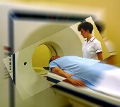
TUESDAY, Nov. 27 (HealthDay News) -- A technique that combines a PET scan with a CT scan can help spot the spread of inflammatory breast cancer, a rare but very aggressive form of the disease, researchers report.
"It is a quicker way of assessing everything," said lead researcher Dr. Selin Carkaci, assistant professor of radiology at The University of Texas M.D. Anderson Cancer Center in Houston. "Our results show it is very useful," she said.
Carkaci was expected to present her findings Monday at the annual meeting of the Radiological Society of North America, in Chicago.
According to the American Cancer Society, IBC accounts for 1 percent to 5 percent of all breast cancer cases in the United States. Often by the time it is discovered, it has spread. The five-year survival rate for those with IBC is 25 percent to 50 percent, according to the cancer society.
The new combo technology has been used in recent years for assessing other cancers as well, Carkaci noted. While the separate technologies of PET (positron emission tomography) and CT (computed tomography) have been in use since the early 1970s, their combined use came into practice only about eight years ago. Patients being treated at cancer centers across the United States should have access to this test, which is becoming more common, Carkaci said.
The technique she used is called FDG-PET/CT -- for "F-18 fluorodeoxyglucose positron emission tomography and computed tomography".
In a PET scan, a small amount of the radioactive drug F-18-labeled glucose is injected into the body. Fast-growing cancer cells feed on sugars and absorb it more quickly than do normal cells, Carkaci explained, so they "light up" on the images created by the PET scanner.
On the other hand, "the CT scanner takes a series of X-ray pictures, which are combined by a computer to create an extremely detailed image of the internal organs or other parts of the body," she said.
The scan gives great anatomical detail including the size, shape and location of the tumor. Together, the tests provide the most complete data on the tumor and its spread, Carkaci said.
In the study, doctors scanned 41 women newly diagnosed with inflammatory breast cancer. They found metastases in 20 patients, or 49 percent. They used biopsy or additional imaging to confirm the results when possible. Biopsy confirmation of metastases were available in four of 13 patients and additional imaging confirmation in nine.
"We found that FDG-PET/CT is 95 percent accurate in identifying distant metastases and 98 percent accurate in identifying regional lymph node metastases," Carkaci said.
"This [scanning] is done immediately after diagnosis, and then following the chemotherapy, to evaluate the response to treatment," she said.
The hope is to stop the cancer in its tracks.
One co-author, Dr. Homer Macapinlac, chair and professor of nuclear medicine at M.D. Anderson, reported being a consultant for General Electric Company and Siemens AG. Both companies produce scanners.
The study findings are "significant for the high rate of metastatic disease, with almost one half of the women with PET/CT able to identify the tumors at an early stage," said Dr. David Bluemke, professor of radiology and medicine and clinical director, MRI, Johns Hopkins Medical Institution, Baltimore.
"The data is very compelling for routine and early use of PET/CT for patients with inflammatory breast cancer," he said. "Although the study was small, the high accuracy rate appears promising for patients with this condition."
More information
To learn more about inflammatory breast cancer, visit the U.S. National Cancer Institute.
Copyright © 2007 ScoutNews, LLC. All rights reserved. 
HealthDayNews articles are derived from various sources and do not reflect federal policy. healthfinder.gov does not endorse opinions, products, or services that may appear in news stories. For more information on health topics in the news, visit the healthfinder.gov health library.