| Table of Contents | ||
| Back Issues | ||
| Search | ||
| Editors | ||
| About FSC | ||
| Instructions for Authors |
| Research and Technology | |
Microscopy of Hair Part II: A Practical Guide and Manual for Animal Hairs |
|
| Douglas W.
Deedrick Supervisory Special Agent Scientific Analysis Section Sandra L. Koch Physical Scientist, Forensic Examiner Trace Evidence Unit Federal Bureau of Investigation Quantico, Virginia |
|
| Introduction
| Basic Structure of Hair | Hair
Identification | Scale Casts | Animal
Hairs Deer Family and Antelope | Commercial Fur Animals | Domestic Animals | Conclusions Report | Testimony | Significance and Value | Glossary | References |
|
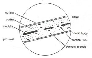
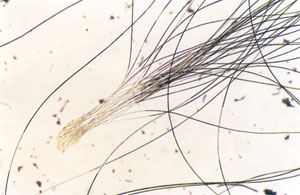
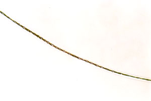
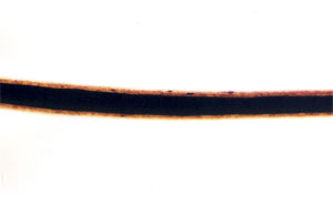
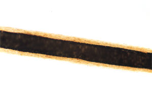
IntroductionDuring the course of a criminal investigation, the identification and comparison of human and animal hairs can be helpful in demonstrating physical contact with a suspect, victim, and crime scene. Hairs can provide investigators with valuable information for potential leads. In Microscopy of Hair Part I: A Practical Guide and Manual for Human Hairs, the characteristics of human hairs were described in detail as well as their significance to an investigation (Deedrick and Koch 2004). In Microscopy of Hair Part II: A Practical Guide and Manual for Animal Hairs, the focus is on animal hairs. This manual is intended to focus on animal hair evidence in the forensic setting and to provide a foundation for its proper identification and comparison. Although Part I and Part II are intended to be a complete manual for the examination of hair, each part can stand alone. Some repetition between the two documents will, therefore, occur because the basics are the same for microscopy of hair, whether human or animal. Basic Structure of HairHair can be defined as a slender, thread-like outgrowth from a follicle in the skin of mammals. Composed mainly of keratin, it has three morphological regions—the cuticle, medulla, and cortex. These regions are illustrated in Figure 82 with some of the basic structures found in them. The illustration is a diagram used to emphasize structural features discussed in this guide. Certain structures may be omitted, and others enhanced for illustrative purposes.
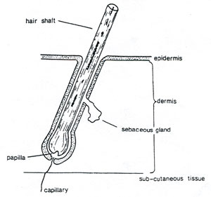
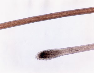
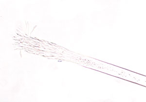
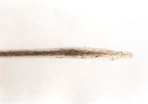
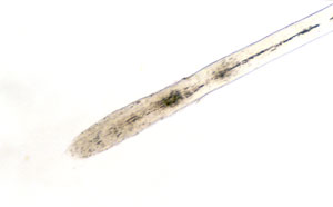
A hair grows from the papilla and, with the exception of that point of generation, is made up of dead, cornified cells. It consists of a shaft that projects above the skin and a root that is imbedded in the skin. Figure 83 diagrams how the lower end of the root expands to form the root bulb. Its basic components are keratin (a protein), melanin (a pigment), and trace quantities of metallic elements. These elements are deposited in the hair during its growth and/or absorbed by the hair from an external environment. After a period of growth, the hair remains in the follicle in a resting stage to eventually be sloughed from the body.
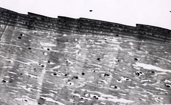
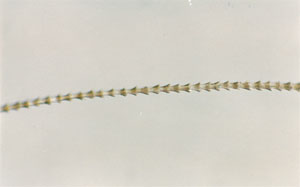
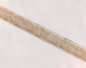
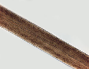
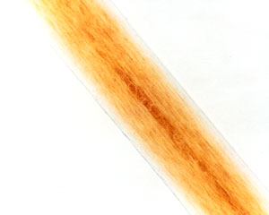
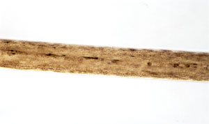
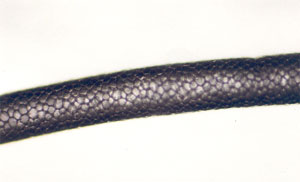
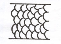
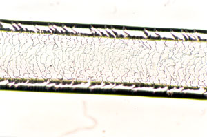
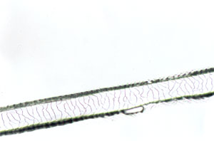
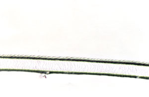
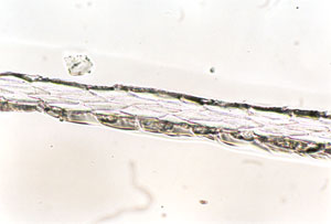
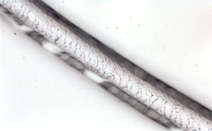
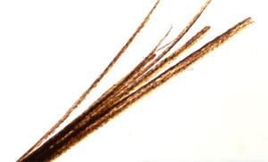
Cuticle The cuticle is a translucent outer layer of the hair shaft consisting of scales that cover the shaft. Figure 84 illustrates how the cuticular scales always point from the proximal or root end of the hair to the distal or tip end of the hair.
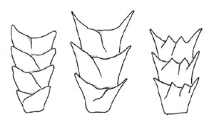
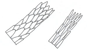
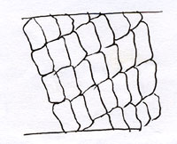
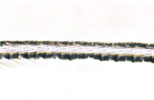
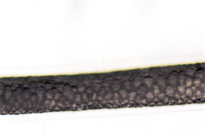
There are three basic scale structures that make up the cuticle—coronal (crown-like), spinous (petal-like), and imbricate (flattened). Combinations and variations of these types are possible. Figures 85-90 illustrate scale structures.
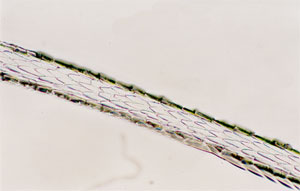
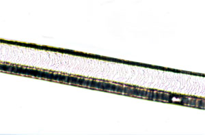
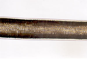
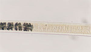
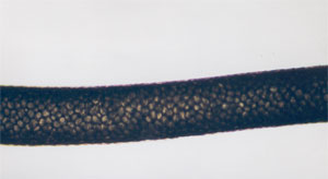
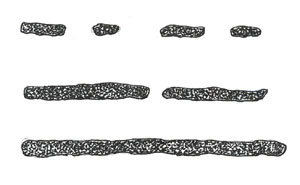
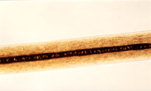
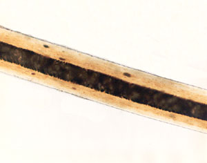
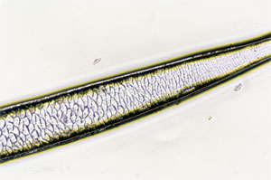
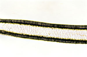
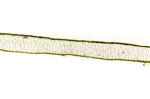
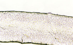
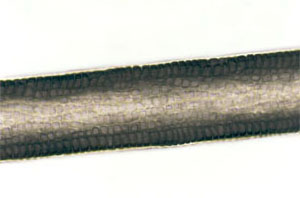
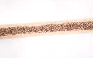
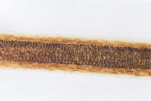
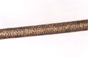
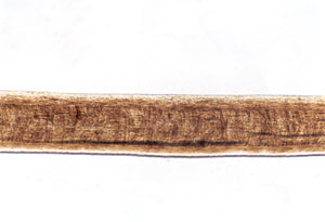
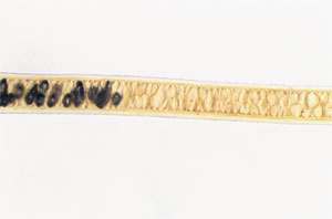
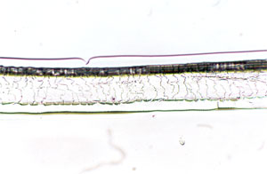
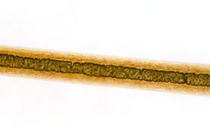
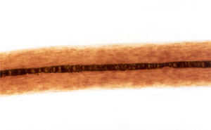
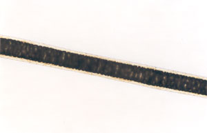
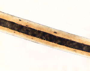
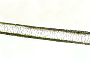
Medulla The medulla is a central core of cells that may be present in the hair. If it is filled with air, it appears as a black or opaque structure under transmitted light, or as a white structure under reflected light. If it is filled with mounting medium or some other clear substance, the structure appears clear or translucent in transmitted light, or nearly invisible in reflected light. In human hairs, the medulla is generally amorphous in appearance, whereas in animal hairs, its structure is frequently very regular and well defined. Figures 91 through 94 are photomicrographs of medullary types found in animal hairs. Figure 91 exhibits a uniserial ladder, and Figure 92 exhibits a multiserial ladder, both found in rabbit hairs. Figure 93 exhibits the cellular or vacuolated type common in many animal hairs. Figure 94 exhibits a lattice found in deer family hairs.
When the medulla is present in human hairs, its structure can be described as fragmentary or trace, discontinuous or broken, or continuous. Figure 95 is a diagram depicting the three basic medullary types.
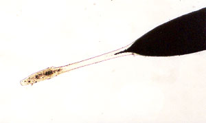
Cortex The cortex is the main body of the hair composed of elongated and fusiform (spindle-shaped) cells. It may contain cortical fusi, pigment granules, and/or large oval-to-round-shaped structures called ovoid bodies. Cortical fusi in Figure 96 are irregular-shaped airspaces of varying sizes. They are commonly found near the root of a mature human hair, although they may be present throughout the length of the hair.
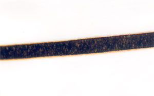
Pigment granules are small, dark, and solid structures that are granular in appearance and considerably smaller than cortical fusi. They vary in color, size, and distribution in a single hair. In humans, pigment granules are commonly distributed toward the cuticle as shown in Figure 97, except in red-haired individuals as in Figure 98. Animal hairs have the pigment granules commonly distributed toward the medulla, as shown in Figure 99.
Ovoid bodies are large (larger than pigment granules), solid structures that are spherical to oval in shape, with very regular margins. They are abundant in some cattle and dog hairs as well as in other animal hairs. To varying degrees, they are also found in human hairs.
Hair IdentificationAnimal Versus Human Hairs Human hairs are distinguishable from hairs of other mammals. Animal hairs are classified into three basic types.
Other types of hairs found on animals include tail hair and mane hair (horse). Human hair is not so differentiated and might be described as a modified combination of the characteristics of guard hairs and fur hairs. Human hairs are generally consistent in color and pigmentation throughout the length of the hair shaft, whereas animal hairs may exhibit radical color changes in a short distance called banding. The distribution and density of pigment in animal hairs can also be identifiable features. The pigmentation of human hairs is evenly distributed, or slightly more dense toward the cuticle, whereas the pigmentation of animal hairs is more centrally distributed, although more dense toward the medulla. The medulla, when present in human hairs, is amorphous in appearance, and the width is generally less than one-third the overall diameter of the hair shaft. The medulla in animal hairs is normally continuous and structured and generally occupies an area of greater than one-third the overall diameter of the hair shaft. The root of human hairs is commonly club-shaped, whereas the roots of animal hairs are highly variable between animals.
The scale pattern of the cuticle in human hairs is routinely imbricate. Animal hairs exhibit more variable scale patterns. The shape of the hair shaft is also more variable in animal hairs. Scale CastsIt may be necessary to make a scale cast of the hair specimen in order to see the scale pattern more clearly, particularly in the identification of some animal hairs. Ogle and Mitosinka (1973) devised a quick and easy method of making a scale cast using a Polaroid film-print coater. A thin layer is applied to a glass microscope slide with two or three passes of the Polaroid print coater. The hair specimen is lightly pressed onto the film and allowed to stand until the film is dry. The hair is then pulled from the film, and the cast remains. A method developed by Crocker (1998) at the Centre of Forensic Sciences in Toronto, Canada, uses clear tape and coverslip together as a mounting medium, which allows for quick observation of such surface features as the scale pattern. Scale casts may also be prepared using clear nail polish. A thin coat is painted on a glass microscope slide or, if the lacquer is thinned with acetone, a drop may be allowed to run down the surface of the slide. The hair to be cast is placed on the slide and allowed to dry. When the surface has dried, the hair is removed to reveal the scale pattern. Animal Hairs IdentificationThe animal hairs discussed in this manual will be limited to those animals most likely to be encountered in casework. An adequate reference collection is essential for accurate identification of questioned specimens. In most cases, specific identification can only be accomplished with guard hair specimens. In some instances, however, as with some commercial garment furs, specimens can be identified on the basis of the microscopic appearance of the fur or down hairs alone. The animal hairs presented here can be classified into three major groups on the basis of their microscopic appearance.
For individual identification in these groups, deer family and antelope hairs are distinguished on the basis of their scale patterns, whereas commercial fur animals are distinguished on the basis of their color, color bands, scale patterns, and medullary structure. Domestic animals are distinguished primarily through their root structure, medullary structure, and pigmentation. Deer Family and AntelopeThe deer family and antelope hairs are not readily distinguishable on the basis of their gross appearance or microscopic appearance in whole mount. However, when scale cast impressions are carefully studied, certain patterns become apparent that can be used to separate the different members of this group. It is emphasized that these patterns are impressions perceived through general observation of the entire hair specimen. Group Characteristics
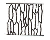
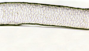
Deer (White-Tailed Deer and Mule Deer) Scale margins are round and isodiametric and resemble fish scales.
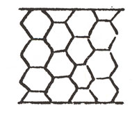
Caribou Scales are hexagonal and usually longer than wide.
Elk Scales are elongated and five- or six-sided. Scale margins are narrow and ends are pointed.
Moose Scales are relatively large with irregular polygons. Overall hair diameter is considerably larger than other members of this group.
Antelope Scales are diamond-shaped and frequently give impression of being arranged in diagonal rows.
Deer family hairs exhibit similarities to goat hairs in general form (medullary structure). However, goat hairs are generally finer in overall diameter (approximately 220μ) and exhibit a narrow, pigmented cortex. No cortex is apparent in deer family hairs (cortex is only occasionally visible in elk hairs). The scale pattern of goat hairs shows flattened scale margins and no regular pattern. Commercial Fur AnimalsThe commercial fur group includes several specimens commonly encountered in fur garments that are distinctive in their microscopic appearance. These are rabbit, seal, mink, muskrat, and chinchilla. Of these, seal and chinchilla are identified on the basis of the appearance of their down or fur hairs, not by their guard hairs. This is because guard hairs are frequently plucked from seal pelts for a more pleasing appearance. Chinchilla pelts have few, if any, guard hairs. The remaining specimens in the commercial fur group are identifiable largely by their characteristic colors and color banding. Group Characteristics
Rabbit: Extensively Used in Felted Fabrics, Glove Linings, Trim, Coats. Sheds Readily.
Seal: Coats
Mink: Coats, Stoles, Hats, Trim
Muskrat: Coats, Trim
Chinchilla: Coats, Hats, Trim
Raccoon (Procyon): Coats, Trim
Red Fox: Stoles, Trim
Beaver: Coats, Hats
Bear (Black and Grizzly): Coats, Rugs
Domestic AnimalsThere are wide variations in color and length of the hair specimens in the domestic animal group. The identifying characteristics given are general and apply in most cases. In order to distinguish between dog and cat and between beef (cattle) and horse, it is usually necessary that the root be present. Group Characteristics
Cat
Dog
Cattle
Horse
Hog
Goat
ConclusionsThere are three basic conclusions that can be reached from a microscopic examination and comparison of hairs.
In the first conclusion, it is stated that the questioned hairs can be associated with the source of the known hairs. Hairs are biological specimens and subject to variation. During the analysis of hair, the examiner must establish the range of variation in the sample and then determine whether the questioned hair fits in that range. The possibility cannot be dismissed that there may be two hair samples whose ranges of variation overlap and distinguishing between the samples is not possible. ReportThe information contained in the report should be limited to a factual statement of findings concerning the examinations conducted. An interpretation of the evidence is saved for court testimony and explanation about the basis for the examinations. The report should be clear, concise, and easily understood. Technical terminology that is foreign to a layperson or contributor does not serve a useful purpose. The report should contain information pertinent to the requests made by the contributor of the evidence and to the examinations performed. A listing of the items of evidence and their origin, either a description or the contributor's reference number, should be included. Results of examinations should be written clearly, followed by a statement of the examiner's conclusions. A statement may follow to clearly state the limiting factors of hair examinations. Example
Hair comparisons are not a means of absolute personal identification. The statement of results sets forth, fairly completely, those determinations that can be made (i.e., that the hairs came from a brown dog, that they exhibit characteristics of a specific breed or animal type, and that the Q hairs are consistent with the K hairs in microscopic appearance). The resulting conclusion is that given these results, the Q hairs can be associated with the suspect's dog Rover or another of similar breed or type. Mitochondrial DNA analysis can be conducted on animal hairs, but it is not currently done in any state or federal forensic laboratory. TestimonyExpert witness testimony should include an education component on hair evidence for the jury and judge and a statement of the results as reported. The witness should be prepared to discuss the procedures used in reaching the conclusion(s) and to defend opinions. An expert witness should endeavor to promote a better understanding of the methods of examination, the theory of the transfer of trace materials, and the strengths and limitations of the science. Significance and ValueThe forensic analysis of hair has been accepted in courts of law for many years, but this does not necessarily validate the science. The reliability of hair examinations must be weighed with the education and training of the examiner, as well as with the procedures used in the analysis. The examinations must be objective and impartial, and the weight placed on the results must be in accordance with the experience and training of the examiner. Hair identifications are subjective interpretations of objective criteria. The variability and distribution of the microscopic characteristics are useful in determining whether or not a questioned hair could have originated from a particular individual. Animal hairs are different in this respect from human hairs. Animal hairs do not posses enough individual characteristics to differentiate individual animals from other similar animals. However, differentiation between breeds and types of animals is possible and may be useful in an investigation. Studies (Bisbing and Wolner 1984; Gaudette and Keeping 1973) have been conducted to determine the significance of hair associations. Some of these studies attempted to establish a mathematical probability of a match. The FBI Laboratory does not use the mathematical calculations of other researchers nor does it support the feasibility of establishing a numerical probability of a hair match. The ability to analyze and interpret hair characteristics is a skill gained by training and testing. New examiners must study hair samples from different animal species and breeds and take hair-matching tests to demonstrate the ability to correctly associate hairs with a particular source. Hair associations in casework should be subject to a confirmation process that has another qualified examiner review the match before a report is issued. The significance of a hair match is dependent on the distinct qualities of the hairs and the experience of the examiner. The greater the number of associations found in casework and the greater the number of cross matches, the greater the chance of association. GlossaryAnagen: Actively growing root stage. Catagen: A transitional root stage between the actively growing anagen stage and the resting telogen stage. Comparison microscope: Two microscopes joined by an optical bridge with a split screen to see both fields of views at the same time. Cortex: Middle portion of hair extending from the cuticle to the medulla and containing the pigment granules, cortical fusi, and ovoid bodies. Cortical fusi: Air spaces located in the cortex of hairs. Cuticle: Translucent outer layer of the hair shaft consisting of overlapping scales. Fur hairs: Fine hairs that make up the undercoat of mammals and provide warmth. Guard hairs: Coarse hairs that provide protection and are usually longer than fur hairs. Keratin: Fibrous protein forming the chemical basis for hair, nails, and feathers. Medulla: Central portion of hair, the core area. Melanin: A pigment that gives hair its color. Miscible: Able to be mixed together. Ovoid bodies: Dark bodies of unknown origin that are a useful discriminatory characteristic in their pattern of appearance. Papilla: Connective tissue from which hair is generated from the follicle. Pigment granules: Melanin granules whose size, shape, density, and distribution vary. Scales: Outermost portion of the cuticle, flattened and imbricate in humans, pointing toward the distal end of the hair shaft. Tactile hair: (Vibrissae): Whiskers only found on animals. Telogen: Resting stage when the root takes on club shape and is ready to be naturally shed. Vellus: Fine body hair. ReferencesBisbing, R. E. and Wolner, M. F. Microscopical discrimination of twins' head hair, Journal of Forensic Sciences (1984) 29(3):780-786. Crocker, E. J. A new technique for the rapid simultaneous examination of medullae and cuticular patterns of hairs, Microscope (1998) 46(3):169-173. Deedrick, D. W. and Koch, S. L. Microscopy of hair Part I: A practical
guide and manual for human hairs, Forensic Science Communications
[Online]. (January 2004). Available: www.fbi.gov/hq/lab/fsc/backissu/jan2004/research/ Gaudette B. D. and Keeping, E. S. An attempt at determining probabilities in human scalp hair comparison, Journal of Forensic Sciences (1974) 19(3):599-606. Ogle, R. R. and Mitosinka, G. T. A rapid technique for preparing hair cuticular scale casts, Journal of Forensic Sciences (1973) 18(1):82-83. |
|