|
|||
|
NIOSH Safety and Health Topic:Chest Radiography |
|
|
|
||||||||
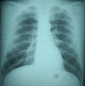
| Radiographic medical techniques have long been a powerful adjunct to disease detection and diagnosis. For many decades, the chest radiograph has been the standard approach to assessing dust diseases of the lung, being relatively simple, inexpensive, non-invasive, and safe. See below for the following topics:
The Role of Classification of Chest Radiographs in Medical DiagnosisAmong those with a history of workplace dust exposure, chest radiographs are part of medical testing procedures for dust-induced lung diseases, including the pneumoconioses (e.g., coal workers’ pneumoconiosis, silicosis, and asbestosis). It should be stressed, however, that although formal classification of the chest radiograph using the ILO system can at times be useful in furthering accurate diagnosis of disease, in general it is not required. As in other settings, it is important to remember that chest radiograph findings alone are insufficient for the diagnosis of pneumoconiosis. Other data, such as the medical and occupational history, the physical examination, additional types of chest imaging, various laboratory tests, and biopsy results should also be considered. Special Considerations for Classification of Chest Radiographs in Medical DiagnosisThe American Thoracic Society (ATS) and other medical organizations publish official guidelines for diagnosis and management of certain respiratory diseases, including asbestosis (ATS 2004). These guidelines emphasize the importance of using multiple diagnostic modalities. Physicians should be aware that dust exposure is not only implicated in the development of the pneumoconioses, but can cause other pulmonary diseases, such as emphysema, COPD, and cancer. In addition, pneumoconiosis may be present with a normal or near-normal chest radiograph. Also, abnormalities consistent with pneumoconiosis may be nonspecific on a standard chest radiograph. Computerized tomography may be helpful in clarifying such cases. Factors Relevant to Classification of Chest Radiographs in Medical Diagnosis1. ILO classification ILO classification is not necessary for medical diagnosis of pneumoconiosis. However, the ILO classification system can be useful in describing occupationally-induced abnormalities, if present. If pneumoconiosis is suspected, an ILO classification may eventually be required for participation in Federal or State compensation systems (see sections below on Government Programs and Contested Proceedings). 2. Remuneration Not applicable to medical diagnosis. 3. Reader selection A B Reader is not required for diagnosis of any pneumoconiosis. 4. Number of readers and summary classification methods Not applicable to medical diagnosis. 5. Blinding The physician providing the clinical interpretation of the chest radiograph need not be blinded to other information pertaining to the patient. In fact, an optimal clinical interpretation takes into account work and exposure history and other medical information concerning the case. 6. Quality assurance Physicians providing the clinical interpretation of the chest radiograph for diagnostic purposes should evaluate their need for participating in quality assurance procedures in relation to their involvement in classifying radiographs for other settings ( worker monitoring, epidemiologic research, government programs, or contested proceedings). 7. Notification Once the diagnosis is made, ethical practice demands that physicians disclose findings to the patient in a timely manner, provide appropriate medical follow-up, educate patients about their illness, and advise them to reduce or eliminate further exposure, as appropriate, in order to prevent progression of the disease. Physicians should be mindful that recognition of occupational lung disease can provide an opportunity for preventive interventions not only for the affected worker but also for the associated workplace, process, agent, or industry. Physicians and other health care providers are encouraged, and in some states required, to notify their State of diagnosed or suspected cases of occupational pneumoconioses, including silicosis and asbestosis. A chest radiograph classified or otherwise interpreted as consistent with the reportable disease is often considered sufficient evidence to require reporting. If physicians are not already aware of their State reporting requirements, they should contact their state to be apprised of any reporting requirements for which they may be responsible. Contacts for State Public Health Departments can be found on the Association for State and Territorial Health Officials (ASTHO) Web site (external link). Physicians should also inform their patients about filing deadlines for state Workers’ Compensation and Federal Black Lung benefits. Patients should be advised that there are often time limits that apply to how long individuals have to make a claim after they are diagnosed with a compensable disease. To further disease identification and to promote prevention, reporting of diagnosed or suspected cases of pneumoconiosis to state public health organizations is required in some states. ReferencesAmerican Thoracic Society. Diagnosis and Initial Management of Nonmalignant Diseases Related to Asbestos. Am J Respir Crit Care Med 2004;170:691-715. Association for State and Territorial Health Officials
Page last updated:
February 6, 2008
Page last reviewed: March 6, 2008 Content Source: National Institute for Occupational Safety and Health (NIOSH) |
|||||||||