|
|||||||||||||||
Sexually Transmitted Diseases > Gonorrhea > Laboratory Information > Biochemical
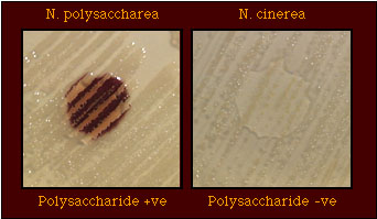
Tests > Polysaccharide from Sucrose Polysaccharide from Sucrose Test PrincipleSome bacterial species produce a starch-like polysaccharide from sucrose which stains dark blue-purple to black with iodine. Among the Neisseria spp., N. perflava biovar perflava, N. mucosa, N. sicca, N. flavescens, and N. polysaccharea produce polysaccharide from sucrose. This test is invaluable for differentiating between strains of N. meningitidis (polysaccharide-negative) and N. polysaccharea (polysaccharide-positive); as many as 25% of organisms identified as nontypable N. meningitidis strains were found to be N. polysaccharea when tested with the polysaccharide production test. Traditionally, polysaccharide production was detected on a starch-free medium containing 5% (w/v) sucrose. However, 5% sucrose is inhibitory for some strains ; polysaccharide may be detected on a starch-free medium containing 1% sucrose. Strains are inoculated onto the medium to give either well-isolated colonies; polysaccharide may not be detected in confluent growth. Although some strains, e.g., N. gonorrhoeae, may not grow on the medium, a polysaccharide test may be performed if a heavy inoculum is deposited on the medium. The test does not required incubation in an a carbon dioxide-supplemented (5%) atmosphere; carbon dioxide may be provided to support growth of the strain. After incubation for 48 h. at 35-36.5C in a incubator with or without supplemental carbon dioxide, the starch-like polysaccharide, if produced, is detected by adding a drop of iodine solution (Gram's iodine or Lugol's iodine [Gram's iodine diluted 1:4]) to the growth; the polysaccharide stains dark blue-purple to black (Figure 1). The growth of strains which do not produce polysaccharide may acquire a light brown color due to the iodine solution but, otherwise, shows no darkening (Figure 1). Figure 1. Production of Polysaccharide from Sucrose by N. polysaccharea, but Not by N. cinerea. N. polysaccharea (left) produces polysaccharide from sucrose; N. cinerea (right) does not produce polysaccharide from sucrose.
Although the traditional method for detection of polysaccharide recommends incubation for 48 h., polysaccharide may be detected after incubation for 24 h. and up to 3-4 days. However, strains that produce the polysaccharide may also degrade it through the action of amylosucrase. Thus, the reaction may be weaker, or absent, if strains are incubated longer than 48 h.; polysaccharide may not be detected if strains are incubated for 5 days. The stain reaction may fade with time but will immediately reappear if more iodine solution is added to the growth. It is not possible to detect polysaccharide in the sucrose-containing medium of rapid acid detection tests such as QuadFERM+. Specimen RequirementsOptimum specimen: Unacceptable specimen: Compromising factors affecting test results:
Stability of specimen:
Reagents & EquipmentMedium: Polysaccharide medium (Tryptic soy agar containing 1% sucrose) Tryptic soy agar (Difco), 40.0 g
Store medium at 4C to 10C (refrigerated) until used. Prewarm the medium to room temperature before inoculation. Reagent: Lugol's iodine solution (Gram's iodine solution diluted 1:4) Note: Iodine solutions provided with commercially available Gram strain kit reagents may not react with polysaccharide to give a positive reaction with strains known to be positive in this test. It may be necessary to make the iodine solution from the original formulation for Gram's iodine. Alternatively, Gram's iodine may be used. Store iodine solution (Gram's iodine) at room temperature (15C to 30C) in the dark (wrapped in aluminum foil). Reagent may be used until quality assurance tests fail. Quality Control/Test ProcedureQC strains:
Problems & SolutionsThe reaction of a polysaccharide-positive strain may fade with time. The reaction may be developed again by adding another drop of iodine to the growth. False-negative reactions may be obtained if inoculated medium is incubated longer than 48 h. because amylosucrase (produced by polysaccharide-positive organisms) degrades the polysaccharide that has been produced. The polysaccharide may not be detected with commercially available Gram's iodine reagent. Limitations of TestIf the test is performed properly and quality control strains give appropriate results, there should be no limitations to this test. Care must be taken to ensure that all components of the test are performed properly. No identification of genus or species may be made on the basis of this test alone. The polysaccharide test aids differentiation among strains that may exhibit identical acid production patterns or produce hydroxyprolylaminopeptidase-positive or gamma-glutamylaminopeptidase-positive reactions in enzyme substrate tests. Results, Interpretation, & ReportingIsolates may be reported as: "Polysaccharide-positive" if the growth of an isolate turns dark brown, purple, or black after the addition of Gram's iodine solution. "Polysaccharide-negative" if the color of the growth does not change color other than the color contributed by the iodine reagent. BibliographyBerger U. Polysaccharidbildung durch saprophytische Neisserien. Zentralbl Bakteriol Parasitenkd Infektionskr Hyg Abt 1 Orig. 1961;183:345-348. Knapp JS. Historical perspectives and identification of Neisseria and related species. 1988. Clin Microbiol Rev 1988;1:415-431. Boquette MT, Marcos C, Saez-Nieto JA. Characterization of Neisseria polysaccharea sp. nov. (Riou, 1983) in previously identified noncapsular strains of Neisseria meningitidis. J Clin Microbiol 1986;23:973-975. Knapp JS, Hook EW III. Prevalence and presistence of Neisseria cinerea and other Neisseria spp. in adults. J Clin Microbiol 1988;26:896-900. |
|||||||||||||||
Content provided by the Division of STD Prevention |
|||||||||||||||
|
|||||||||||||||