Your lungs are organs in your chest that allow your
body to take in oxygen from the air. They also help remove carbon dioxide (a
waste gas that can be toxic) from your body.
The lungs' intake of oxygen and removal of carbon
dioxide is called gas exchange. Gas exchange is part of breathing. Breathing is
a vital function of life; it helps your body work properly.
Other organs and tissues also help make breathing
possible. (For more information, see "The
Respiratory System.")
The Respiratory System
The respiratory system is a group of organs and
tissues that help you breathe. The main parts of this system are the airways,
the lungs and linked blood vessels, and the muscles that enable breathing.
The Respiratory System
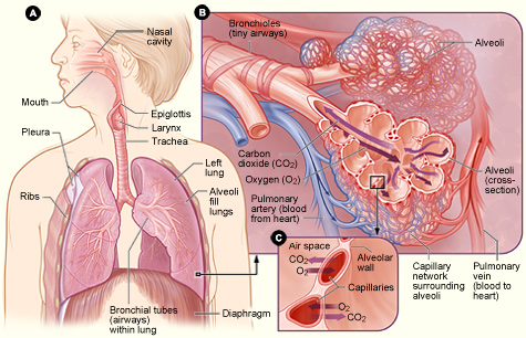
Figure A shows the location of the
respiratory structures in the body. Figure B is an enlarged image of airways,
alveoli, and the capillaries. Figure C shows the location of gas exchange
between the capillaries and alveoli.
Airways
The airways are pipes that carry oxygen-rich air to
your lungs and carbon dioxide, a waste gas, out of your lungs. The airways
include your:
Nose and linked air passages called nasal
cavities
Mouth
Larynx (LAR-ingks), or voice box
Trachea (TRA-ke-ah), or windpipe
Tubes called bronchial tubes or bronchi, and
their branches
Air first enters your body through your nose or
mouth, which wets and warms the air. (Cold, dry air can irritate your lungs.)
The air then travels through your voice box and down your windpipe. The
windpipe splits into two bronchi that enter your lungs.
A thin flap of tissue called the epiglottis
(ep-i-GLOT-is) covers your windpipe when you swallow. This prevents food or
drink from entering the air passages that lead to your lungs.
Except for the mouth and some parts of the nose, all
of the airways have special hairs called cilia (SIL-e-ah) that are coated with
sticky mucus. The cilia trap germs and other foreign particles that enter your
airways when you breathe in air.
These fine hairs then sweep the particles up to the
nose or mouth. There, they're swallowed, coughed, or sneezed out of the body.
Nose hairs and mouth saliva also trap particles and germs.
Lungs and Blood Vessels
Your lungs and linked blood vessels deliver oxygen
to your body and remove carbon dioxide. Your lungs lie on either side of your
breastbone and fill the inside of your chest cavity. Your left lung is slightly
smaller than your right lung to allow room for your heart.
Within the lungs, your bronchi branch into thousands
of smaller, thinner tubes called bronchioles. These tubes end in bunches of
tiny round air sacs called alveoli (al-VEE-uhl-eye).
Each of these air sacs is covered in a mesh of tiny
blood vessels called capillaries. The capillaries connect to a network of
arteries and veins that move blood through your body.
The pulmonary (PULL-mun-ary) artery and its branches
deliver blood rich in carbon dioxide (and lacking in oxygen) to the capillaries
that surround the air sacs. Inside the air sacs, carbon dioxide moves from the
blood into the air. Oxygen moves from the air into the blood in the lungs.
The oxygen-rich blood then travels to the heart
through the pulmonary vein and its branches. The heart pumps the oxygen-rich
blood out to the body. (For more information on blood flow, see the Diseases
and Conditions Index
"How
the Heart Works" article.)
The lungs are divided into five main sections called
lobes. Some people need to have a diseased lung lobe removed. However, they can
still breathe well using the rest of their lung lobes.
Muscles Used for Breathing
Muscles near the lungs help expand and contract
(tighten) the lungs to allow breathing. These muscles include the:
Diaphragm (DI-a-fram)
Intercostal muscles
Abdominal muscles
Muscles in the neck and collarbone area
The diaphragm is a dome-shaped muscle located below
your lungs. It separates the chest cavity from the abdominal cavity. The
diaphragm is the main muscle used for breathing.
The intercostal muscles are located between your
ribs. They also play a major role in helping you breathe.
Beneath your diaphragm are abdominal muscles. These
help you breathe out when you're breathing fast (for example, during physical
activity).
Muscles in your neck and collarbone area help you
breathe in when other muscles involved in breathing don't work properly, or
when lung disease impairs your breathing.
What Happens When You Breathe?
Breathing In (Inhalation)
When you breathe in, your diaphragm contracts
(tightens) and moves downward. This increases the space in your chest cavity,
into which your lungs expand. The intercostal muscles between your ribs also
help enlarge the chest cavity. They contract to pull your rib cage both upward
and outward when you inhale.
As your lungs expand, air is sucked in through your
nose or mouth. The air travels down your windpipe and into your lungs. After
passing through your bronchial tubes, the air finally reaches and enters the
alveoli (air sacs).
Through very thin walls of the alveoli, oxygen from
the air passes to the surrounding capillaries (blood vessels). A red blood cell
protein called hemoglobin (HEE-muh-glow-bin) helps move oxygen from the air
sacs to the blood. (Oxygen is especially drawn to hemoglobin.)
At the same time, carbon dioxide moves from the
capillaries into the air sacs. The gas has traveled in the bloodstream from the
right side of the heart through the pulmonary artery.
Oxygen-rich blood from the lungs is carried through
a network of capillaries, which become the pulmonary vein. This vein delivers
the oxygen-rich blood to the left side of the heart. The left side of the heart
pumps the blood to the rest of the body. There, the oxygen in the blood moves
from blood vessels into surrounding tissues.
(For more information on blood flow, see the
Diseases and Conditions Index
"How
the Heart Works" article.)
Breathing Out (Exhalation)
When you breathe out, your diaphragm relaxes and
moves upward into the chest cavity. The intercostal muscles between the ribs
also relax to make the chest cavity size smaller.
As the chest cavity gets smaller, air rich in carbon
dioxide is forced out of your lungs and windpipe, and then out of your nose or
mouth.
Breathing out requires no effort from your body
unless you have a lung disease or are doing physical activity. When you're
physically active, your abdominal muscles contract and push your diaphragm even
more so against your lungs. This pushes the air in your lungs out rapidly.
The animation below shows how the lungs work. Click
the "start" button to play the animation. Written and spoken explanations are
provided with each frame. Use the buttons in the lower right corner to pause,
restart, or replay the animation, or use the scroll bar below the buttons to
move through the frames.
The animation shows how the lungs
inhale oxygen and transfer it to the blood. It also shows how carbon dioxide (a
waste product) is removed from the blood and exhaled.
What Controls Your Breathing?
A respiratory control center at the base of your
brain controls your breathing. This center sends ongoing signals down your
spine and to the nerves of the muscles involved in breathing.
These signals ensure your breathing muscles contract
(tighten) and relax regularly. This allows your breathing to happen
automatically, without you being aware of it.
To a limited degree, you can change your breathing
rate, such as by breathing faster or holding your breath. Your emotions also
can change your breathing. For example, being scared or angry can affect your
breathing pattern.
Your breathing will change depending on how active
you are and the condition of the air around you. For example, you need to
breathe more often when you do physical activity. In contrast, your body needs
to restrict how much air you breathe if the air contains irritants or
toxins.
To adjust your breathing to changing needs, your
body has a number of sensors in your brain, blood vessels, muscles, and
lungs.
Sensors in the brain and in two major blood vessels
(the carotid (ka-ROT-id) artery and the aorta) detect carbon dioxide or oxygen
levels in your blood and change your breathing rate as needed.
Sensors in the airways detect lung irritants. The
sensors can trigger sneezing or coughing. In people who have
asthma,
the sensors may cause the muscles around the airways in the lungs to contract.
This makes the airways smaller.
Sensors in the alveoli (air sacs) detect a buildup
of fluid in the lung tissues. These sensors are thought to trigger rapid,
shallow breathing.
Sensors in your joints and muscles detect movement
of your arms or legs. These sensors may play a role in increasing your
breathing rate when you're physically active.
Lung Diseases and Conditions
Many steps are involved in breathing. If injury,
disease, or other factors affect any of the steps, you may have trouble
breathing.
For example, the fine hairs (cilia) that line your
upper airways may not trap all of the germs you breathe in. These germs can
cause an infection in your bronchi (bronchitis) or deep in your lungs
(pneumonia). These infections cause a buildup of mucus and/or fluid that
narrows the airways and hinders airflow in and out of your lungs.
If you have
asthma,
breathing in certain substances that you're sensitive to can trigger your
airways to narrow. This makes it hard for air to flow in and out of your
lungs.
Over a long period, breathing in cigarette smoke or
air pollutants can damage the airways and the air sacs. This can lead to a
condition called
COPD
(chronic obstructive pulmonary disease). COPD prevents proper airflow in and
out of your lungs and can hinder gas exchange in the air sacs.
An important step to breathing is the movement of
your diaphragm and other muscles in your chest, neck, and abdomen. This
movement lets you inhale and exhale. Nerves that run from your brain to these
muscles control their movement. Damage to these nerves in your upper spinal
cord can cause breathing to stop, unless a machine is used to help you breathe.
(This machine is called a ventilator or a respirator.)
A steady flow of blood in the small blood vessels
that surround your air sacs is vital for gas exchange. Long periods of
inactivity or surgery can cause a blood clot called a
pulmonary
embolism to block your lung artery. This reduces or stops the flow of blood
in the small blood vessels and interferes with gas exchange.
Key Points
Your lungs are organs in your chest that allow
your body to take in oxygen from the air. They also help remove carbon dioxide
(a waste gas that can be toxic) from your body.
The respiratory system is a group of organs and
tissues that help you breathe. The main parts of this system are the airways,
the lungs and linked blood vessels, and the muscles that enable breathing.
The airways are pipes that carry oxygen-rich
air to your lungs and remove carbon dioxide from your lungs.
Your lungs and linked blood vessels deliver
oxygen to your body and remove carbon dioxide.
Muscles near the lungs expand and contract
(tighten) to allow breathing. These muscles include the diaphragm, intercostal
muscles, abdominal muscles, and muscles in the neck and collarbone area.
When you breathe in, your diaphragm and
intercostal muscles contract to increase the space in your chest cavity, into
which your lungs expand. As your lungs expand, air is sucked in through your
nose or mouth. The air travels down your windpipe and into your lungs' air
sacs.
In the air sacs, oxygen moves from the air into
the blood in the lungs. At the same time, carbon dioxide moves from the blood
in the lungs into the air in the air sacs. Surrounding blood vessels carry the
oxygen-rich air to the rest of the body.
When you breathe out, your diaphragm and
intercostal muscles relax to make the size of the chest cavity smaller. As the
chest cavity gets smaller, air rich in carbon dioxide is forced out of your
lungs and windpipe, and then out of your nose or mouth.
Your breathing is controlled by the base of your
brain and sensors located in the brain, blood vessels, muscles, and lungs.
These sensors adjust your breathing to changing needs.
Many steps are involved in breathing. If injury,
disease, or other factors affect any of the steps, you may have trouble
breathing.