You are here:
Research

Light Imaging Section
Evelyn Ralston, Ph.D.
Section Chief, Light Imaging Section
Office of Science and Technology (OST)
Phone: (301) 402-6479
Fax: (301) 402-3417
E-mail: ralstone@mail.nih.gov
Mission Statement
The Light Imaging Section functions as a Core Facility. It offers NIAMS scientists access to state-of-the-art light imaging equipment and expertise in light imaging techniques. The Facility offers training and assistance in a variety of techniques of light microscopy and digital image analysis.
For all practical questions regarding available equipment, access, as
well as for help to plan your light imaging experiments, NIAMS staff
please check
the link, Core
Facility Information.
Research Overview
Research in the Section focuses on the cell biology of skeletal muscle. Skeletal muscle is made of uniquely organized large multinucleated cells, built through successive developmental changes in cell morphology and subcellular organization. We are currently particularly interested in the organization of the Golgi complex (GC). One of the important functions of the GC is the targeting of membrane proteins. This is an essential function in a cell with distinct membrane domains. Muscle fibers have several membrane domains: the plasma membrane, the T-tubules, the neuromuscular junction, the myotendinous junctions, to which proteins must be specifically targeted. Some of these domains extend at large distances from the nuclei. Instead of compact Golgi complexes associated with the nuclei, muscle fibers have thousands of small Golgi complexes dispersed throughout the fibers. We have shown that their distribution is fiber type-specific and regulated by the patterned contractile activity of the muscle fiber. Although the organization of the GC in muscle appears specific to this tissue, we have shown that it relies on principles common to all mammalian cells linking the Golgi complex, the microtubule cytoskeleton and the endoplasmic reticulum (ER).
Our present work focuses on understanding how the GC changes are regulated during muscle development and are caused by contractile activity. Experiments in progress include the relative localization of GC, ER exit sites and centrosomal proteins by immunofluorescence, immunogold EM, and by observation of GFP-tagged proteins in live cells, as well as investigations of the signaling pathway(s) involved. We are currently investigating whether microtubule stabilization, which takes place during muscle differentiation, plays a role in the relocation of the GC. We are also using transgenic mice lacking cytoskeletal proteins to probe their role.

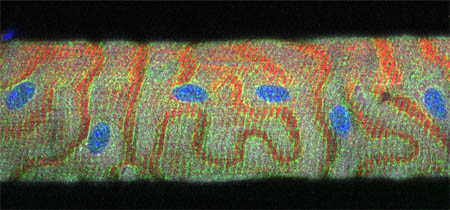
As part of our research on muscle cytoskeleton, a single fiber from a mouse Tibialis Anterior muscle was stained with antibodies against the proteins syne (cytoskeleton) and caveolin-3 (cell surface), and counterstained with Hoechst, a fluorescent dye that labels nuclei. The fiber was photographed in the Zeiss LSM510 confocal microscope. This image is the superposition of a transmitted light image of the fiber (white) and of the immunofluorescence images (red for syne, green for caveolin-3, blue for the nuclei). The blood vessels that surrounded the muscle fiber in the muscle have been removed but have left ditches that appear as sinuous red channels.
Selected Publications
Ralston, E., Lu Z., Biscocho, N., Soumaka, E., Mavroidis, M., Prats, C., Lømo, T., Capetanaki, Y. & Ploug, T. Blood vessels and desmin control the positioning of nuclei in skeletal muscle fibers. J. Cell. Physiol. Published Online: 13 Sep 2006 DOI: 10.1002/jcp.20780.
Bugnard E, Zaal KJ, Ralston E. Reorganization of microtubule nucleation during muscle differentiation. Cell Motil Cytoskeleton. 2005; 60(1): 1-13.
![]()
Ralston, E., Ploug, T., Kalhovde, J., and Lømo, T. Golgi complex, ER exit sites and microtubules in skeletal muscle fibers are organized by patterned activity. J. Neurosci. 2001; 21, 875-883.
Lu Z, Joseph D, Bugnard E, Zaal KJ, Ralston E. Golgi complex reorganization during muscle differentiation: visualization in living cells and mechanism. Mol Biol Cell. 2001; 12:795-808.
![]()
Ralston E, Lu Z, Ploug T. The organization of the Golgi complex and microtubules in skeletal muscle is fiber type dependent. J. Neurosci. 1999; 19:10694-705
![]()
See complete list of publications
Updated December 27, 2007


