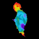
Caption: Novel biosensor system maps the timing and location of Rac protein activation in a living mouse embryo fibroblast. Courtesy of Chris Welch, Lou Hodgson, and Klaus Hahn, University of North Carolina at Chapel Hill.
Video [AVI, 2.95MB]
NIGMS is committed to furthering the development of imaging technologies that can be applied to basic research in cell biology. The Institute is particularly interested in advances that will enable, as an ultimate goal, the visualization of single molecules and single molecular events within living cells.
Visualization at the cellular and subcellular level has led to many fundamental breakthroughs in our understanding of how cell structure and function are related and dynamically regulated. NIGMS has supported many of these advances and will continue to support creative and promising research in this area.
Learn about current NIGMS funding opportunities and previously funded programs in cellular imaging by following the links below.