     |
< Previous | Home | Next >
Color Plates Plate 1: Trichomonads TOP  Trichomonads in a saline wet mount (high power) (Monif, 1982; Fig. 22-2; reprinted with permission from Harper & Row.) Plate 2: Clue cells of G. vaginalis TOP 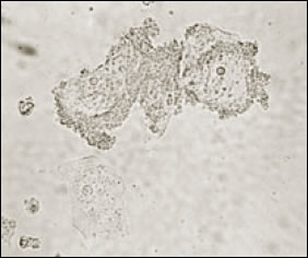 Clue cells of G. vaginalis vaginitis on a saline wet mount (high power) (Monif, 1982; Fig. 22-3; reprinted with permission from Harper & Row.) Plate 3: Candida TOP 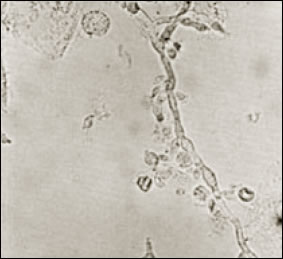 Candida in a saline wet mount (high power) (Monif, 1982; Fig. 22-4B; reprinted with permission from Harper & Row.) Plate 4: Vaginal candidiasis TOP 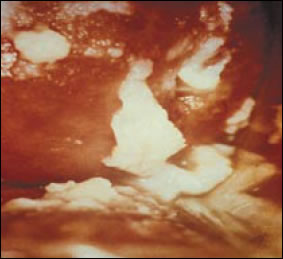 Vaginal candidiasis: thrush patches on the vaginal wall of a patient with candidiasis (courtesy J. Anderson, MD). Plate 5: Severe vulvar intraepithelial neoplasia (VIN3) TOP  Severe vulvar intraepithelial neoplasia (VIN3) (Wilkinson and Stone, 1995; Fig 6.27; reprinted with permission from Williams & Wilkins.) Plate 6: Nontender chancres (kissing lesions) TOP 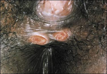 Nontender chancres (kissing lesions) in a woman with primary syphilis (Wilkinson and Stone, 1995; Fig 8.46; reprinted with permission from Williams & Wilkins.) Plate 7: Extensive vulvar condylomata acuminata TOP 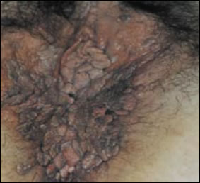 Extensive vulvar condylomata acuminata (human papillomavirus) (Wilkinson and Stone, 1995; Fig 9.3; reprinted with permission from Williams & Wilkins.) Plate 8: Cervical intraepithelial neoplasia (CIN3) TOP  Cervical intraepithelial neoplasia (CIN3) demonstrating coarse mosaicism and punctuation on posterior lip (Burghardt, 1991; Fig. 11.37; reprinted with permission from Thieme Medical Publishers.) Plate 9: Mucopurulent cervicitis TOP 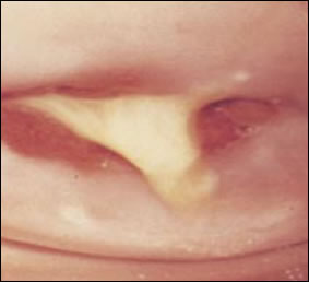 Mucopurulent cervicitis caused by C. trachomatis (Holmes, 1999; Plate 16; reprinted with permission from McGraw Hill.) Plate 10: Profuse purulent frothy vaginal discharge TOP 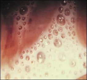 Profuse purulent frothy vaginal discharge due to trichomonas (Holmes, 1999; Plate 11: Pelvic inflammatory disease TOP 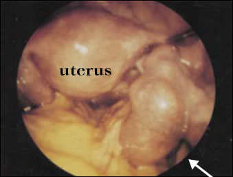 Pelvic inflammatory disease, proven chlamydial pyosalpinx. Right tube is swollen and tortuous (arrow) (Holmes, 1999; Plate 17; reprinted with permission from McGraw Hill.) Plate 12: Chancroid TOP  Chancroid (Holmes, 1999; Plate 32; reprinted with permission from McGraw Hill.) Plate 13: Condyloma latum in secondary syphilis TOP 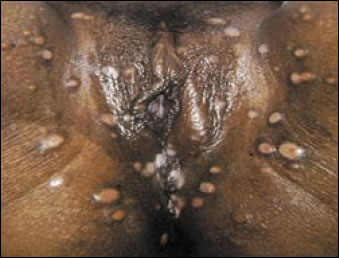 Condyloma latum in secondary syphilis (Holmes, 1999; Plate 47; reprinted with permission from McGraw Hill.) Plate 14: Lesion of herpes simplex TOP 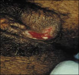 Lesion of herpes simplex (courtesy J. Anderson, MD). Plate 15: Herpes simplex in woman with AIDS TOP 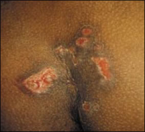 Herpes simplex in woman with AIDS, CD4<50 (courtesy J. Anderson, MD). Plate 16: Apthous genital ulceration TOP 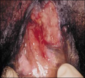 Apthous genital ulceration (courtesy J. Anderson, MD). Plate 17: Apthous oral ulceration TOP  Apthous oral ulceration (courtesy J. Anderson, MD). |
||||||||||||||||||||||||||||||||||||||||||||||||||||||||||
![]()
Top
| Home | HRSA
| HHS |
Disclaimer | Accessibility
| Privacy | Download
Adobe Reader| | Freedom of Information
Act