 |
Glossary of Radiation Physics for Radiation TherapySeveral radioactive sources of beta-particle and gamma-ray emitting radionuclides are currently under investigation for use in brachytherapy. This interdisciplinary field draws on experimental and theoretical nuclear and medical physics. We define here several terms in three areas: i) the nuclear physics of radioactive decay, ii) experimental radiation dosimetry, and iii) theoretical radiation dosimetry. Nuclear Physics of Radioactive DecayRadioactivity is the phenomenon of emissions of neutral or charged particles, or electromagnetic radiations from unstable atomic nuclei (radionuclides). Radioactivity is an amount of a radionuclide in a particular energy state at a given time. Mathematically, it is defined as the quotient of dN by dt, where dN is the number of spontaneous nuclear transformations from that energy state in the time interval dt.The unit of activity in the International System (SI) of units is the becquerel (Bq), which is equal to the unit reciprocal second (s-1). In many fields the older unit, the curie (Ci), is still in use, where 1 Ci = 3.7 × 10¹⁰ Bq (exactly).
The activity of an amount of radionuclide is given by the product of the decay
constant, The half life is the time necessary for one half of the nuclei to decay. The activity at any time t can be computed using the initial activity A0 and the decay time t according to A = A0 exp(-0.693 15 t/T1/2) .
Beta particles are high-energy electrons emitted by the
nucleus in nuclei that contain too many or too few neutrons. For a system with
excess neutrons, the neutron is transformed into a proton and in the process an
electron and an antineutrino 
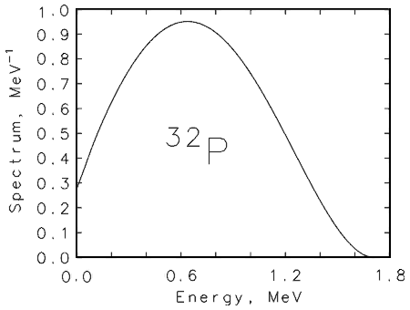
The emitted negatively charged beta particles, usually denoted as
Positrons are positively charged electrons (antiparticles of the
electron) emitted by the nucleus. They are usually denoted as
Gamma rays are photons emitted during nuclear de-excitation processes. These gamma-ray transitions may be from a metastable excited state, or between levels in a daughter nucleus. The large majority of gamma rays from fission-product and man-made radionuclides have energies between 20 keV and 2 MeV. X rays are photons emitted during atomic relaxation processes. x rays are often emitted from radionuclides, however, because orbital electrons are affected by the high-energy processes in the nearby nucleus. In the electron capture process, for example, the nucleus captures an electron (usually a K-shell electron since it is closer to the nucleus), and a proton and electron form a neutron. This process leaves a K-shell vacancy, and a characteristic x ray from the daughter nucleus can be emitted as orbital electrons from higher shells fill the vacancy. Annihilation radiation is a form of photon radiation associated with a class of radioactive decays. Positrons, since they are antiparticles of ordinary electrons, cannot survive long in normal matter. Thus, positrons emitted during radioactive decay will slow down in matter until they reach thermal equilibrium. They combine with an electron in an annihilation event in which their combined mass (1.022 MeV) is converted to energy. This takes the form of two annihilation quanta of 0.511 MeV each which are oppositely directed (to insure the conservation of momentum).
Bremsstrahlung is the photon radiation emitted by the
deceleration of an electron in the Coulomb field of an atom. Thus,
bremsstrahlung radiation is present during all beta decay processes as the
emitted Electromagnetic radiation (gamma-ray and x-ray photons) interacts with matter principally by three processes: photoelectric absorption, Compton scattering and pair production. In the photoelectric process, which dominates at lower energies, the photon transfers its energy to an atomic electron, which is then ejected from the atom with an energy equal to that of the incident photon minus the electron's binding energy.
In Compton scattering (incoherent scattering), the photon loses a fraction of its energy to an atomic electron, and a scattered secondary photon emerges generally in a direction different to that of the incident photon. Higher energy photons willundergo multiple Compton scatter events until the process is finally terminated by a photoelectric absorption. For photons with energies exceeding 1.022 MeV, the process of pair production can occur, whereby an electron-positron pair is formed. The positron produced will ultimately annihilate with the production of two 0.511 MeV photons (or in flight). The three interaction processes compete as a function of photon energy, electron density and nuclear charge of the stopping material. Quantitative measures of the photon interactions in matter are attenuation coefficients based on cross sections for specific interactions. The total, narrow-beam attenuation coefficient µ is given by the sum µ = µphotoelectric + µCompton + µpair production. The attenuation process for a beam of photons traversing a slab of matter is an exponential function of the form I = I0 exp(-µ l) ,
where I and I0 are the intensities of the transmitted
beam and the incident beam, respectively, l is the distance travelled in
matter, and µ is the linear
attenuation coefficient. A useful procedure is to express distances in terms of
the mass thickness - the product of density
I = I0 exp[(-µ/
where µ/
The two quantitative parameters used to describe the interaction of
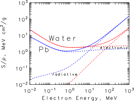
Total mass stopping power, S/
 = 1/
= 1/ ×
(dE/dl)col + 1/ ×
(dE/dl)col + 1/ × (dE/dl)rad
. × (dE/dl)rad
.Range for electrons of a given energy in a material is an important parameter in designing radiation sources. A given particle, however, does not travel in a straight line as it slows down, so the range by its nature is an average value given by
where E is the incident electron energy.

Experimental Radiation Dosimetry
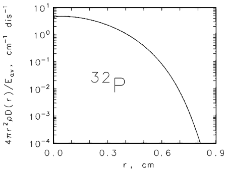
Absorbed dose is the quantity of main interest to the clinician
for both Depth dose defines the relationship between the absorbed dose and depth in tissue. Thus, one can define a reference depth in terms of the absorbed dose at 1 cm tissue depth for photons or 2 mm tissue depth for beta particles. Air kerma strength is the quantity used to specify the strength of a gamma-ray emitting radionuclide. Kerma, kinetic energy released per unit mass, is a measure of the energy released in a volume of air at some distance from a radioactive source. For photon emitting sources used in brachytherapy it has units of Gy s-1 m2. Ionization chambers are the class of radiation detectors in which the radiation produces ionization events in a gas. The gas is contained in a chamber equipped with two electrodes which differ in potential by several hundred to a thousand volts. Ion pairs formed by the incident radiation travel to the positively and negatively charged electrodes. The charge collected (or current) is a measure of the strength of the external radiation source.
Extrapolation chamber is a primary standard chamber used at the NIST to
establish the surface absorbed dose rate for
Well ionization chambers are sealed, pressurized ionization chambers (also called dose calibrators) which are intended for use in assaying brachytherapy sources. Their response must be determined for each source type and will in general depend on the particular source jig and catheter used for the measurement. In-air ionization chambers are chambers which communicate with the air. Thus, humidity, pressure and temperature corrections must be applied. They are widely used for high activity sources, such as high dose rate iridium-192 sources. Scintillation detectors are made of materials that absorb ionizing radiation and convert a fraction of the energy into light. These photons are transmitted by lightguides to photodetectors, which include diodes and photomultiplier tubes. Thermoluminescent dosimeters (TLD) are based on inorganic materials which store a fraction of the energy deposited by an external source. The stored energy may be released by heating the TLD with a thermal source or a laser. Traditional TLDs are too large for use with intravascular therapy seeds, but sheets of material are now available with pixel sizes of 100 µm. Radiochromic films consist of a thin emulsion on a mylar backing in which a chemical detector produces a blue color which is proportional to the amount of radiation received. These films are widely used to measure autoradiographs of seeds used in intravascular brachytherapy. With proper calibration they can also be used to establish the absorbed dose in a phantom material. Phantom is the material used for the absorbing medium in the experimental dose measurement. Several different phantom materials are used in intravascular therapy including water phantoms and those fabricated from plastics (A-150 plastic, tissue-equivalent plastic, solid water).
Theoretical Radiation DosimetryBoth theoretical and computation methods in radiation dosimetry have advanced rapidly over the past two decades. At this point in the development of intravascular radiation therapy they are indispensable tools for seed designers and clinical investigators. The ability to predict the depth dose relationship in a variety of arteries, with different materials (blood, calcifications, smooth muscle cells, etc.), for many radionuclides, and in many configurations (catheters, stents, marker seeds) dictates the use of computational methods in conjunction with experiments to establish the basic dosimetry. Some of the main tools are listed here.
Fermi spectrum refers to the Monte Carlo methods are those computer models based on "chance." A geometry is defined comprising a source and detector medium. Electrons and/or photons are "emitted" from the source and then "followed" and "scored" as they undergo interactions in the medium. Since it is a statistical process, one must have a large number of events (10 million), and the accuracy will also depend on the detailed specification of the source geometry and the medium (lumen, catheter, tissue). ETRAN is a general-purpose coupled electron-photon Monte Carlo transport code developed at the NIST. A related code is the ITS code developed at Sandia National Laboratory which is a more user-friendly code for intravascular radiation therapy calculations. MCNP is a Monte Carlo code developed at Los Alamos National Laboratory which was originally intended for neutrons, but has been extended to include gamma rays and electrons as well. EGS4 is a Monte Carlo code developed at Stanford University and more recently extended to a variety of applications in medical physics. Point kernels for photons or electrons in a specified medium such as water are the results of an analytical calculation of the dose assuming a point source in a homogeneous medium. They are much faster than MC methods but are limited to a few configurations.

ReferencesR.D. Evans, The Atomic Nucleus, New York: McGraw Hill, 1955. G. Choppin, J. Rydberg, J.O. Liljenzin, Radiochemistry and Nuclear Chemistry, 2nd Edition, Oxford: Butterworth-Heinemann, 1995. W.R. Leo, Techniques for Nuclear and Particle Physics Experiments, Berlin: Springer-Verlag, 1992. G.F. Knoll, Radiation Detection and Measurement, 2nd Edition, New York: Wiley & Sons, 1989. Radiation Quantities and Units, Report 33, International Commission on Radiation Units and Measurements, Bethesda: ICRU/NCRP Publications, 1980. A Handbook of Radioactivity Measurements Procedures, Report No. 58, 2nd Edition, National Council on Radiation Protection and Measurements, W.B. Mann (ed), Bethesda, NCRP Publications, 1985. Return to top | Ionizing Radiation Division 
 Inquiries or comments:
bert.coursey@nist.gov.
Online: April 1998 - Last update: July 2001 |