
|
|
High technology health care is one of the leading industry sectors in the world's leading economies. The major killers and debilitating diseases of an aging population are cancer, heart disease, stroke, diabetes, arthritis and neurological disorders. Industry is spending hundreds of millions of dollars on research on new diagnostic and therapeutic tools. The NIST Physics Laboratory plays a major role in developing both research tools and national measurement standards that support U.S. industry and allow our industries to compete, to gain, and to maintain market share in this intense international competition. Medical Physics
Mammography
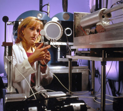
calibration facility Ionizing radiation together with surgery and chemotherapy comprise the main tools in treatment of cancers. Radiation is used in three very different ways, and each of these has a quite separate set of requirements for measurements and standards. Beams of photons and charged particles from accelerators are used to treat about 600,000 patients annually in the U.S. NIST develops detectors such as absorbed dose water calorimeters to measure radiation beams and transfers these calibrations to secondary laboratories including the University of Wisconsin-Madison, K&S Associates in Nashville, the M.D. Anderson Cancer Center in Houston, and the Memorial Sloan Kettering Cancer Center in New York.  Balloon angioplasty © Jeffrey Aarons The second form of radiation treatment is brachytherapy, which is one of the fastest growing markets in radiation devices today. In this application, small radioactive sources are implanted or inserted into the body and the short range gamma-ray and beta-particle radiations kill cancerous cells while minimizing damage to normal tissues. NIST has taken a leading role in developing measurement standards and protocols for these new devices. The current medical research is focused on prostate cancer and prevention of restenosis following balloon angioplasty. However, there are many new radionuclides and devices proposed for this field of therapy and medical centers are considering optimum treatments for other cancers and diseases using these tiny sources. 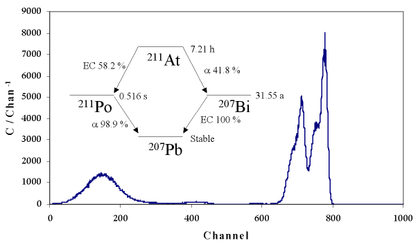
Decay scheme and liquid scintillation spectrum of astatine-211. Radiopharmaceutical Standardization Laboratory The third form of radiation delivery is systemic therapy in which a radioactive labeled pharmaceutical is administered that seeks out the target cells. NIST has worked closely with the pharmaceutical industry of North America to identify new drug products that will be used in clinical trials. Rapid development of NIST standards for new products is an essential first step towards FDA approval for use in humans. Recent advances in targeting cancer cells with monoclonal antibodies, antibody fragments and smaller proteins is driving our research towards beta-particle and alpha-particle emitters that will deliver a high radiation dose to the tumor volume. Biophysics
Terahertz spectroscopy A program is underway to explore the low frequency intramolecular dynamics of proteins and DNAs. Current efforts focus on obtaining THz spectra of models for proteins (e.g., N-methylacetamide) and small, synthetic DNA oligomers (e.g., poly(A)4). We then plan to employ mid-infrared and far-infrared (THz) time-resolved spectroscopies to directly monitor low frequency, concerted motions of small proteins and helical DNA oligomers or related systems. Such measurements will extract protein-folding rates and determine mechanisms responsible for DNA base pair hydrogen-bonding, surface interactions and helix dynamics. These investigations use state-of-the-art pulsed THz generation and detection methods including GaAs antennas and ZnTe nonlinear crystals for broadband spectroscopic determinations and imaging of short-chain DNA probes on supports. Application of molecular dynamics modeling and 2D correlation techniques are also being employed for identifying molecular motions responsible for observed THz spectra. Enhanced Raman spectroscopy Raman spectroscopy is being applied to the conformational studies of small peptides in crystals, in rare-gas matrixes, and in solution. In addition, we are using polarized Raman spectroscopy to determine the secondary and tertiary structures of membrane proteins and their orientation with respect to the membrane. In these studies, the proteins are bound to synthetic lipid bilayers or bicelles and aligned in a high magnetic field for study. The alignment is similar to what can be done with liquid crystals with electric or magnetic fields and has been successfully used in bimolecular NMR spectroscopy. By studying these aligned proteins with polarized Raman spectroscopy we obtain additional data about the orientation of the bond-polarizability tensors with respect to the known polarization direction of the laser. This information is combined with molecular models to infer details about the structure of the protein. The Raman spectrometer presently consists of a single-frequency Ar+ laser, operating at various frequencies between 455 nm and 514 nm, a He-Ne laser operating at 633 nm, or a single or frequency doubled Ti:Sapphire laser, with tunable frequency output from 700 nm to 975 nm and from 350 nm to 490 nm. The Raman-scattered light is analyzed with a 0.5 cm-1 resolution triple-grating monochromator. The selectivity of the monochromator is sufficient for resolving features to within 10 cm-1 of the excitation frequency. Near field scanning optical microscopy Single molecule probes have been used by others to study structure and dynamics of single proteins in a biological or biomimetic environment. In the next year we plan to extend these studies to include the behavior of single molecules in bioengineered materials. As part of this effort we are constructing an instrument capable of fast full-field single-molecule imaging whose applications include studying translational diffusion. Traditionally, molecular diffusion has been studied in biological systems using fluorescence fluctuation correlation spectroscopy (FCS), a confocal technique. There is some concern that in FCS the diffusion of molecules is affected by the confocal beam. We plan to combine our single-molecule full-field imaging apparatus with a confocal beam to help elucidate the effect of light-forces on single fluorescent molecules in embedded in biological membranes. Electron paramagnetic resonance (EPR) spectroscopy Oxidative and radiation damage to biological tissues result in formation of free radicals and these paramagnetic centers can be quantified by EPR spectroscopy. NIST is one of the leaders in applying this technique to measurement of low levels of radiation doses in bone, tooth enamel and dentin. In a collaboration with Russian scientists and the National Cancer Institute we are developing measurement methods to determine the radiation doses to residents near major nuclear facilities in the old Soviet Union. Research is focused on improving the sensitivity and accuracy to the level that the method can be a quantitative tool in radiation epidemiology. The weak signal from the irradiated hydroxy apatite is confounded by signals from other organic free radicals, and sample preparation techniques and instrumentation must be substantially improved to measure environmental doses. |

 Inquiries or comments:
Feedback
Inquiries or comments:
Feedback